A broken bone is painful and often follows significant trauma. Dogs and cats with fractures may also have injuries to the chest, abdomen, head, spine, or soft tissues that are less obvious than the damaged limb. Safe transport, stabilization, diagnostic imaging, and a patient-specific repair plan are the first priorities.
Fracture treatment can involve a cast or splint, external fixation, plates and screws, pins, wires, or other methods. The best option depends on the bone, fracture pattern, age, size, soft-tissue injury, overall health, and how well the repair can be protected during recovery.
Signs of a Possible Fracture
- Sudden severe limping or refusal to bear weight
- Swelling, pain, or bruising
- An abnormal angle, shortening, or rotation of the limb
- A dangling or unstable limb
- An open wound near the painful area
- Bone visible through a wound
- Crying, panting, trembling, hiding, or inability to settle
- Difficulty standing or walking after trauma
Some fractures are not visibly deformed. A pet may still walk on an incomplete, stable, or small fracture. An examination and imaging are needed to distinguish a fracture from a joint dislocation, ligament injury, muscle injury, or other source of pain.
When a Fracture Is an Emergency
Seek prompt care after a vehicle accident, major fall, crushing injury, animal attack, or other significant trauma. Go immediately if the pet has breathing difficulty, pale gums, collapse, uncontrolled bleeding, severe distress, neurologic changes, an open fracture, or multiple injuries.
COVE’s Emergency Medicine and Critical Care team can assess and stabilize trauma patients. Orthopedic repair may follow after life-threatening concerns are addressed.
Safe Transport After an Injury
Keep movement to the minimum needed for safety and transport. Use a carrier for a small pet or a firm, flat surface for a larger patient when practical. A painful animal may bite even when normally gentle, so keep hands away from the mouth and avoid unnecessary handling.
Do not pull, straighten, or repeatedly examine the limb. Do not apply a homemade splint or tight bandage unless a veterinary professional provides specific instructions. An incorrectly placed splint can worsen pain, pressure, swelling, or tissue damage.
Do not give human pain relievers or leftover medication. Call the receiving veterinary facility for transport guidance when possible.
How Fractures Are Diagnosed
Whole-Patient Assessment
The team first evaluates breathing, circulation, pain, neurologic status, and other trauma concerns. Bloodwork, chest or abdominal imaging, ultrasound, or monitoring may be needed before definitive orthopedic treatment.
Orthopedic Examination
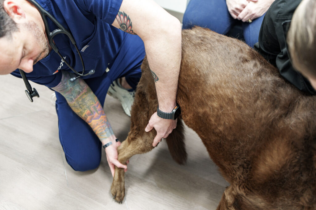
The veterinarian assesses the limb, skin, blood supply, nerve function, joints above and below the injury, and soft-tissue damage. Open wounds are examined carefully because contamination and loss of blood supply can affect treatment and healing.
Radiographs and Advanced Imaging
Radiographs show fracture location, pattern, displacement, joint involvement, and bone quality. Multiple views are usually required, often with sedation or anesthesia for accurate positioning and comfort. CT may be recommended for complex, joint, pelvic, or spinal fractures and for three-dimensional planning.
Fracture Patterns and Why They Matter
Fractures may be complete or incomplete, simple or fragmented, closed or open, and located within a bone shaft, near a joint, through a growth plate, or directly in a joint surface. The pattern affects stability, blood supply, repair options, and recovery.
Young animals can heal more quickly, but growth-plate injuries may alter limb growth. Older pets or patients with other disease may heal more slowly. Open fractures require management of contamination and soft-tissue injury in addition to bone stabilization.
Fracture Treatment Options
Splints and Casts
External coaptation may be appropriate for selected fractures below the elbow or knee when the joints above and below can be immobilized. It requires frequent monitoring because swelling changes, pressure sores, moisture, slipping, and loss of alignment can occur.
Upper-limb fractures are difficult to immobilize effectively with a splint because the shoulder or hip cannot be included. Surgery is often considered when a fracture is unstable, displaced, involves a joint, or cannot be controlled externally.
Plates and Screws
Plates and screws can provide rigid internal stabilization and allow precise alignment. They are commonly used for many long-bone, joint, and complex fractures. Implant selection depends on bone size, fracture forces, and repair strategy.
Pins, Wires, and Nails
Intramedullary pins, wires, interlocking nails, or combinations may be used for selected patterns. Each controls different forces, so combinations or another method may be necessary.
External Skeletal Fixation
Pins placed through the skin into bone connect to an external frame. This can be useful for selected open fractures, soft-tissue wounds, or patterns where an external construct offers practical advantages. Pin sites and the frame require careful home management.
Joint and Growth-Plate Fractures
Fractures involving a joint surface often need accurate reduction to support joint function and limit uneven loading. Growth-plate fractures may require timely stabilization to preserve alignment and growth potential. The surgeon will explain realistic risks, including arthritis or altered growth.
What Happens on Surgery Day?
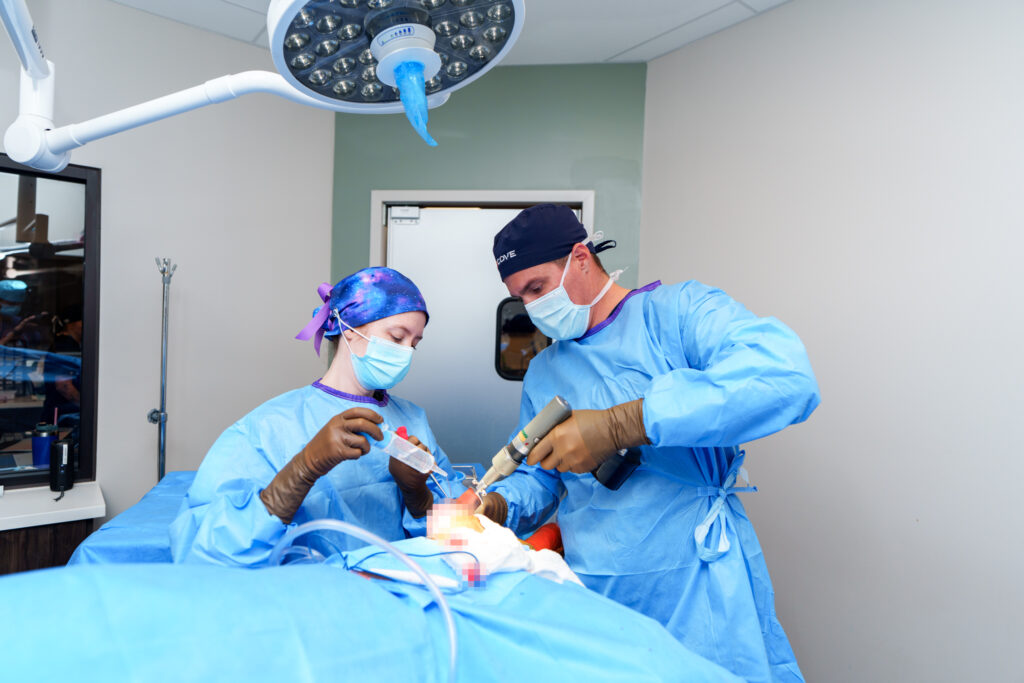
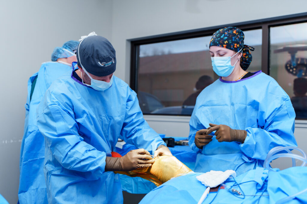
The patient receives anesthesia, monitoring, pain management, and surgical preparation appropriate to health and injury. The surgeon aligns and stabilizes the fracture while protecting blood supply and soft tissues. Postoperative radiographs assess the repair. Hospitalization depends on trauma, comfort, mobility, and medical needs.
Recovery at Home
Activity restriction is central to fracture healing. The plan may include crate or room confinement, nonslip flooring, leash-only bathroom breaks, a support sling, incision or bandage care, prescribed medication, and separation from other pets.
A pet may begin using the limb before the fracture is fully healed. Do not allow running, jumping, furniture access, stairs, or rough play until radiographs and examination support progression. The patient’s discharge instructions always override a general timeline.
Bandage and Splint Monitoring
Keep bandages clean and dry. Call if they slip, become wet or soiled, smell unusual, feel tight, or if toes become swollen, cold, painful, or discolored. Never remove or replace a medical bandage unless instructed.
Rechecks and Radiographs
Rechecks evaluate the incision, bandage or frame, limb use, comfort, and alignment. Follow-up radiographs show whether the fracture is healing before activity increases or implants are removed.
Possible Complications
Complications can include infection, incision or wound problems, delayed healing, failure to heal, loss of alignment, implant irritation or failure, joint stiffness, muscle loss, arthritis, and nerve or blood-vessel injury. Excessive activity can cause a setback even when the pet appears comfortable.
Contact the team for worsening pain, swelling, discharge, odor, a damaged bandage, sudden loss of limb use, fever, low appetite, or any significant fall or escape. Seek emergency care for breathing difficulty, collapse, uncontrolled bleeding, or severe distress.
Fracture Care at COVE
COVE Pet Care evaluates and treats orthopedic trauma for dogs and cats in Columbus, Clintonville, Delaware, and across Central Ohio. Emergency and surgical teams coordinate stabilization, imaging, repair planning, hospital care, and recovery guidance.
Explore orthopedic surgery at COVE, review the guide to limping and orthopedic warning signs, or contact COVE Pet Care. Major trauma and suspected open fractures should be evaluated promptly.