Patellar luxation occurs when a dog’s kneecap moves out of the groove where it normally tracks. It may cause a brief skipping gait, recurring back-leg lameness, pain, or progressive changes in limb alignment. Some dogs have no obvious symptoms, while others need surgical correction.
The condition is also called a luxating patella, slipped kneecap, medial patellar luxation (MPL), or lateral patellar luxation (LPL). The grade is useful, but treatment is not chosen by grade alone. Symptoms, age, anatomy, progression, arthritis, and other knee problems all matter.
How the Kneecap Normally Works
The patella sits within the quadriceps mechanism and normally glides in a groove at the end of the femur. The patellar tendon attaches to the tibial crest below the knee. Together, these structures help straighten the knee and keep the limb aligned during movement.
With patellar luxation, the kneecap moves to the inside or outside of the groove. Medial luxation means it moves toward the inside of the limb; lateral luxation means it moves toward the outside.
Why Patellar Luxation Happens
Some patellar luxations follow trauma, but many are developmental. The problem can involve the alignment and shape of the entire limb rather than only the kneecap. A shallow femoral groove, rotation or angulation of the femur or tibia, an off-center tibial crest, and soft-tissue imbalance may contribute.
Small breeds are commonly affected, but large dogs can also develop medial or lateral patellar luxation. Both knees may be involved. Because conformation and heredity can play a role, owners should discuss breeding implications with their veterinarian.
Symptoms of a Luxating Patella
- Skipping for several steps and then walking normally
- Briefly carrying a back leg
- Shaking or extending the leg before using it again
- Recurring lameness after activity
- Stiffness, pain, or reluctance to jump
- A bow-legged or knock-kneed appearance
- Continuous lameness in more advanced cases
- Reduced activity or difficulty rising
A skipping gait is suggestive but not diagnostic. CCL disease, hip problems, arthritis, muscle injury, and other conditions can look similar. The guide to common causes of limping in dogs explains additional warning signs.
Patellar Luxation Grades
Grade 1
The kneecap can be moved out of the groove during examination but returns to its normal position. Some dogs have no symptoms or only occasional skipping.
Grade 2
The kneecap moves out of place intermittently and may remain luxated until the leg is extended or the patella is repositioned. Repeated movement can contribute to cartilage wear, inflammation, and more frequent lameness.
Grade 3
The kneecap is out of the groove most of the time but can be moved back during examination. Limb-alignment changes are often more apparent, and function may be consistently affected.
Grade 4
The kneecap remains permanently luxated and cannot be returned to the groove during examination. Significant bone and soft-tissue changes may be present.
Grades describe patellar position and stability, not the dog’s entire clinical picture. A low-grade luxation can be painful, while another dog with a higher grade may compensate differently. Surgical decisions require a full orthopedic assessment.
How Patellar Luxation Is Diagnosed
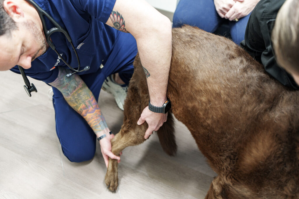
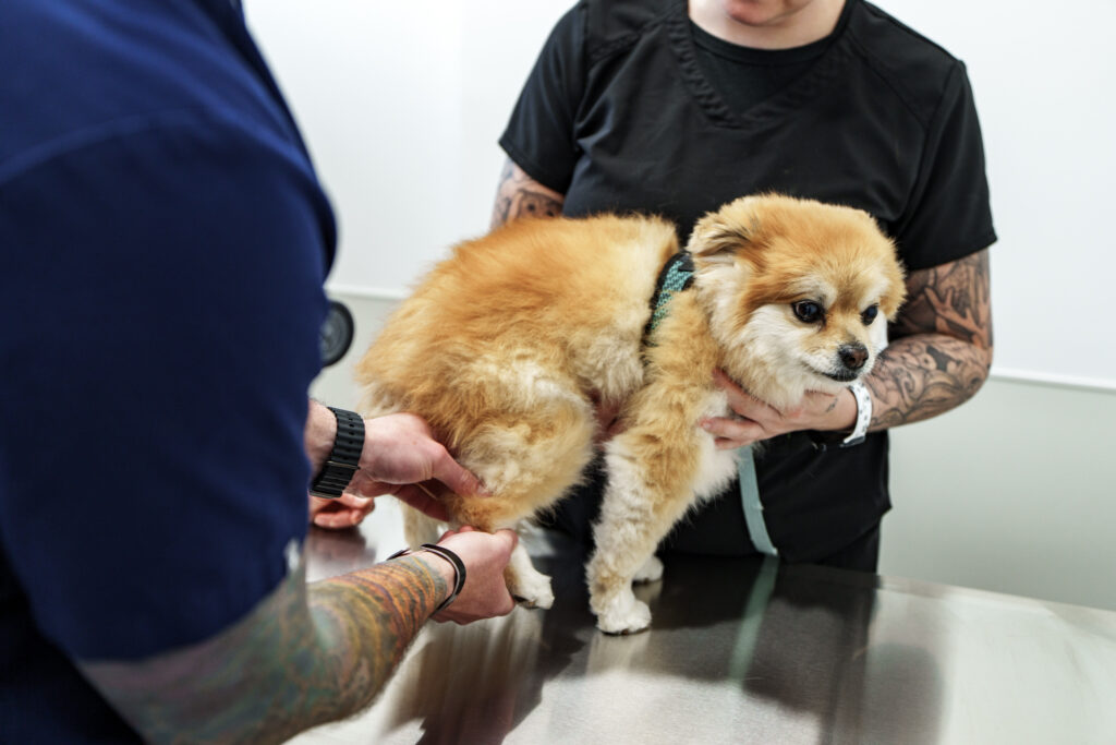
Diagnosis is based primarily on palpation of the kneecap during an orthopedic examination. The veterinarian assesses which direction it luxates, how easily it moves, whether it returns, the range of motion, pain, muscle mass, gait, and stability of other knee structures.
Radiographs help evaluate the pelvis, femur, knee, and tibia for arthritis, bone shape, alignment, and other disease. CT may be recommended when complex femoral or tibial deformity requires three-dimensional planning. Sedation can improve examination and imaging accuracy in selected dogs.
When Monitoring May Be Appropriate
An incidental, low-grade luxation that causes no pain or functional limits may be monitored. The plan may include healthy weight management, appropriate exercise, and rechecks for changes. Owners should watch for more frequent skipping, persistent lameness, reduced activity, or pain.
Monitoring does not mean ignoring the knee. Progression, growth, arthritis, or a second knee problem can change the recommendation.
When Surgery May Be Considered
Surgery may be discussed when patellar luxation causes repeated or persistent lameness, pain, cartilage damage, progressive deformity, or meaningful loss of function. Growing dogs with severe limb-alignment changes may need timely assessment.
No single operation fits every luxating patella. The surgeon selects one or more techniques based on the structures contributing to instability.
Common Surgical Techniques
Soft-Tissue Balancing
Tissues may be released on the side pulling the patella out of position and tightened on the opposite side. This helps center the kneecap but may not be enough when bone alignment is abnormal.
Trochlear Groove Procedures
The femoral groove may be deepened while preserving joint cartilage so the patella can track more securely.
Tibial Crest Transposition
The tibial crest and attached patellar tendon can be moved to improve alignment of the quadriceps mechanism. Pins or other implants hold the bone while it heals.
Corrective Osteotomy
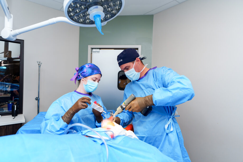
When the femur or tibia has substantial angulation or rotation, the surgeon may cut and realign the bone and secure it with a plate and screws. Complex cases often require detailed imaging and planning.
Patellar Luxation and CCL Disease
Patellar instability can coexist with cranial cruciate ligament disease. A dog may have skipping from the kneecap and pain or instability from a partial or complete CCL tear. The team needs to identify which problems are clinically important and whether they should be addressed together.
Pet owners often call CCL disease an ACL tear. Review the TPLO and CCL surgery guide for an explanation of knee instability, meniscal injury, and treatment options.
Recovery After Patellar Luxation Surgery
Recovery depends on the procedures performed. Bone work requires activity restriction while healing occurs. Instructions may include confinement, leash-only bathroom trips, incision protection, prescribed medication, controlled exercise, rehabilitation, and follow-up radiographs.
A dog may feel energetic before bone and soft tissues are ready for normal forces. Running, jumping, stairs, and rough play should not resume until the surgical team approves them. The patient’s discharge instructions always override a general online timeline.
Possible Risks and Long-Term Care
Possible complications include infection, incision problems, implant irritation or failure, delayed bone healing, fracture, recurrence of luxation, persistent lameness, and arthritis. Risk depends on severity, anatomy, technique, recovery control, and other patient factors.
Surgery aims to improve alignment and function, but it cannot remove all existing arthritis or guarantee a particular outcome. Healthy weight, appropriate exercise, rehabilitation, and ongoing joint care may remain important.
Orthopedic Evaluation at COVE
COVE Pet Care evaluates patellar luxation and other causes of back-leg lameness for families in Columbus, Clintonville, Delaware, and across Central Ohio. The team examines the whole limb, reviews imaging, and explains whether monitoring, rehabilitation, or surgery may be appropriate.
Explore orthopedic surgery at COVE or request a consultation. Online information cannot determine the grade, cause, or best treatment for an individual dog’s kneecap instability.