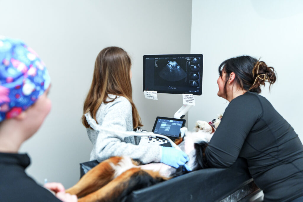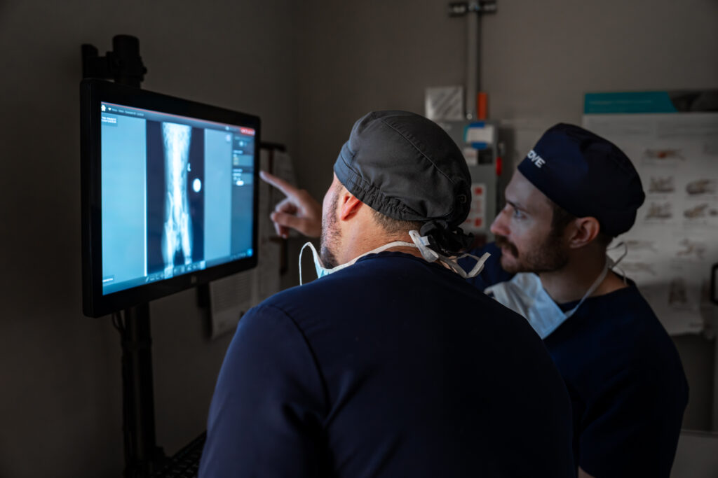
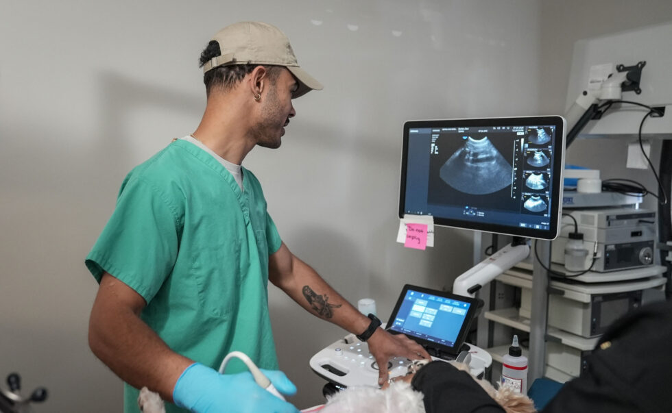
Veterinary X-rays, also called radiographs, create two-dimensional images of structures inside a dog or cat. They are especially useful for evaluating bones, joints, the chest, the abdomen, and the overall size or position of organs. An X-ray may answer the clinical question by itself, or it may help the care team decide whether ultrasound or another test is needed.
At COVE Pet Care, X-rays may be performed for emergency, hospitalized, or specialty patients when the veterinary care team recommends them. They are not offered as a directly scheduled, stand-alone outpatient service.
What are veterinary X-rays?
During an X-ray study, a small amount of ionizing radiation passes through the body and is captured by a digital detector. Dense structures such as bone appear lighter, while air-filled areas such as healthy lungs appear darker. Fat, fluid, and soft tissues appear in shades of gray.
Because a radiograph flattens a three-dimensional body into a two-dimensional image, structures can overlap. That is why the team often takes two or more views from different positions. The exact number depends on the body area, the suspected condition, and whether additional angles are needed.
What can X-rays show in dogs and cats?
Radiographs can reveal changes in size, shape, position, density, and alignment. They may identify a fracture, joint change, abnormal lung pattern, enlarged organ, bladder stone, gas pattern, or a foreign material that blocks X-rays. The image is only one part of the evaluation, however. Your veterinarian interprets it alongside your pet’s symptoms, medical history, physical examination, and laboratory results.
Bone and joint X-rays
X-rays are commonly used to evaluate fractures, dislocations, arthritis, bone infection, some bone lesions, and changes in joint alignment. Precise positioning matters. A painful pet may need medication or sedation so the team can obtain diagnostic views without causing unnecessary stress or movement.
Some soft-tissue injuries around a joint do not appear clearly on radiographs. Tendons, ligaments, cartilage, and subtle early changes may require a different imaging study or follow-up examination.
Chest X-rays
Chest radiographs can help evaluate the lungs, airways, heart silhouette, ribs, diaphragm, and space around the lungs. They may be recommended for coughing, breathing changes, trauma, fever, suspected fluid, or concern about disease spreading to the chest.
An X-ray can show that the heart silhouette is enlarged, but it cannot evaluate heart function in the same way as an echocardiogram. A heart ultrasound is a separate study and is not the same as a routine abdominal ultrasound.
Abdominal X-rays
Abdominal radiographs provide an overview of the stomach, intestines, liver, spleen, kidneys, bladder, and surrounding structures. They can help evaluate vomiting, abdominal pain, urinary concerns, trauma, abnormal distention, stones, and a possible intestinal obstruction.
If a pet may have swallowed an object, some materials are visible directly, including certain metals, stones, and dense bones. Fabric, plastic, wood, and other materials may be difficult to see. In those cases, the veterinarian may recognize secondary signs such as an unusual gas pattern or distended bowel. Read more about foreign body obstruction in dogs and cats.
What may not appear clearly on an X-ray?
Radiographs do not show every disease or swallowed object. Many soft tissues have similar shades of gray, small abnormalities can be hidden by overlapping anatomy, and microscopic disease may not change an organ’s appearance. Timing also matters: early disease may be less visible than changes seen later.
When the X-ray does not fully answer the question, the care team may recommend repeat views, blood or urine testing, ultrasound for dogs and cats, tissue sampling, or another appropriate diagnostic test. Our guide to X-ray versus ultrasound explains how the two studies complement each other.
Will my dog or cat need sedation?
Many pets can have X-rays while awake with gentle positioning. Sedation may be recommended when a pet is painful, anxious, unable to remain still, or needs a carefully positioned view. It can reduce repeated handling and help the team obtain useful images safely. The decision is individualized based on the study, the pet’s condition, and medication or anesthesia risks.
How long do pet X-rays take?
Image acquisition itself may be brief, but the full process can take longer. The team must review the clinical question, prepare and position the patient, obtain the required views, check image quality, and arrange interpretation. Emergency needs, pain control, sedation, additional views, or other tests can affect timing. Your care team can give the most useful estimate for your pet’s situation.
What should owners expect?
The veterinarian will explain why radiographs are recommended and what body area needs to be examined. Owners generally are not present in the X-ray area because staff follow radiation-safety procedures. After the study, the veterinarian reviews the images and connects the findings to the rest of the medical evaluation.
A board-certified veterinary radiologist has advanced training in obtaining and interpreting medical images. Specialist review can be valuable when findings are subtle, the case is complex, or the treatment plan depends on a detailed interpretation. At COVE, radiographs taken during emergency visits are reviewed by radiologists to support the care plan.
When are X-rays urgent?
Seek prompt veterinary care if your pet has trouble breathing, has collapsed, cannot stand, has severe pain, experiences major trauma, repeatedly retches without producing vomit, has a swollen abdomen, cannot urinate, or may have swallowed a dangerous object. Imaging may be part of the emergency evaluation, but stabilization and examination come first. COVE’s Emergency Medicine and Critical Care service is available for urgent concerns in Central Ohio.
Frequently asked questions
Are X-rays painful for pets?
The X-ray itself is not painful. Positioning may be uncomfortable for a pet with an injury or painful condition, so the team may adjust handling, provide pain relief, or recommend sedation.
How many X-ray views will my pet need?
Most studies require more than one view because each radiograph is a two-dimensional projection. The number depends on the body region and the clinical question.
Can an X-ray diagnose cancer?
An X-ray may reveal a mass, bone change, enlarged organ, or pattern that raises concern, but imaging alone often cannot determine whether a change is cancer. Cytology, biopsy, laboratory testing, or additional imaging may be needed.
Can every swallowed object be seen?
No. Metal and other dense materials may be obvious, while cloth, plastic, wood, or thin materials can be difficult to see. Veterinarians also look for indirect signs of obstruction and may recommend ultrasound, repeat imaging, or surgery when concern remains high.
Can I schedule only an X-ray at COVE?
COVE does not present X-rays as a directly scheduled stand-alone outpatient service for pet owners. Radiographs may be performed for emergency, hospitalized, or specialty patients when recommended by the veterinary care team. Call COVE if you are unsure which care pathway fits your pet.
Talk with COVE about diagnostic imaging
Learn more about Veterinary Radiology and Diagnostic Imaging at COVE. For questions about a current COVE patient, specialty consultation, outpatient ultrasound, or referral records, contact COVE Pet Care.
This article is educational and is not a substitute for an examination or diagnosis by a veterinarian. If your pet is ill, injured, or worsening, contact a veterinary professional promptly.