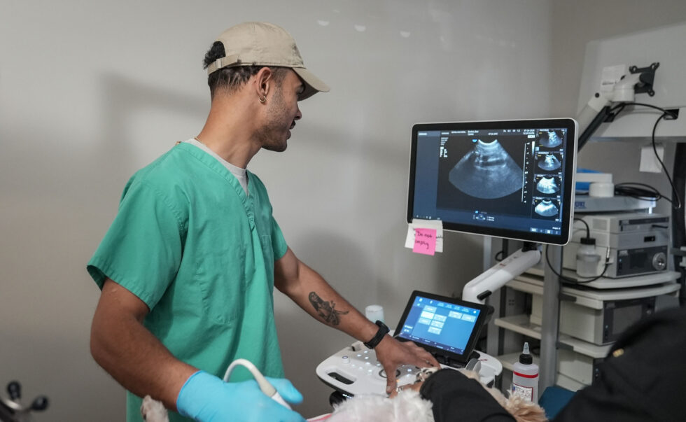
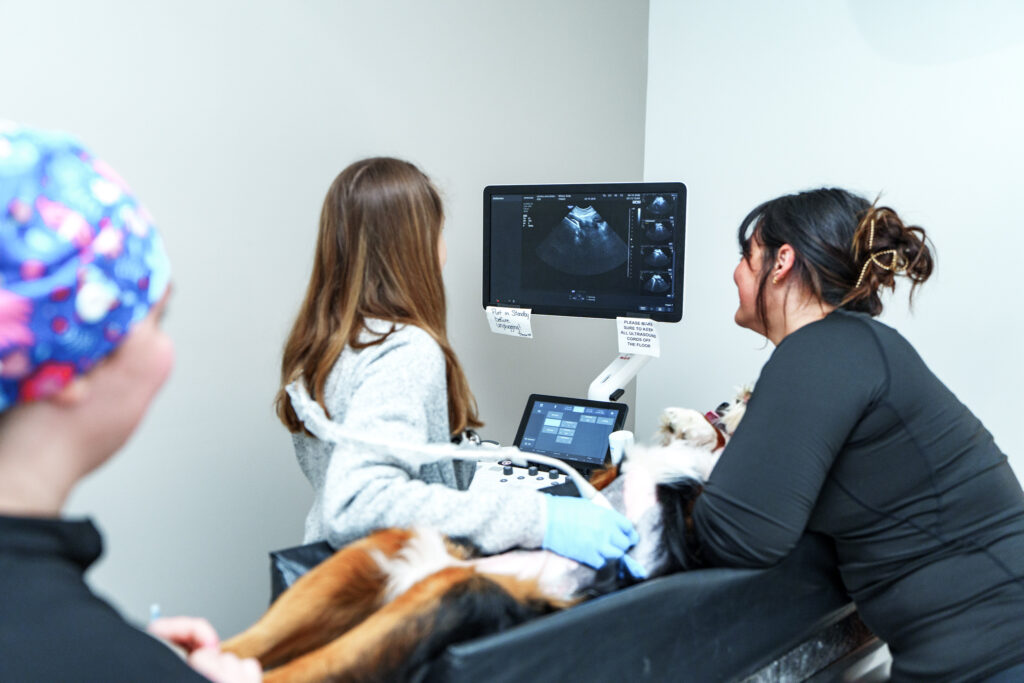
Veterinary ultrasound uses sound waves to create real-time images of structures inside a dog’s or cat’s body. It is noninvasive and does not use ionizing radiation. Veterinarians commonly recommend it when an examination, laboratory test, or X-ray suggests a soft-tissue problem that needs a closer look.
An ultrasound can provide valuable information, but it does not diagnose every condition by itself. Findings are interpreted alongside your pet’s symptoms, examination, laboratory tests, and other imaging.
What can a veterinary ultrasound examine?
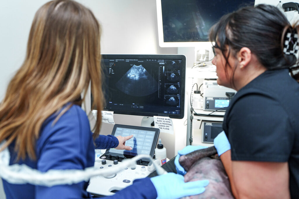
Abdominal ultrasound is often used to evaluate the liver, gallbladder, spleen, kidneys, adrenal glands, urinary bladder, pancreas, lymph nodes, and portions of the stomach and intestines. It can also identify abnormal fluid, changes in organ size or texture, some masses, and signs that a structure is inflamed or obstructed.
Beyond the abdomen, a focused scan may help identify fluid, evaluate a soft-tissue area, or guide a needle. Ultrasound of the heart is called an echocardiogram and is a separate examination.
Common reasons ultrasound is recommended
Your veterinarian may recommend an ultrasound after abnormal bloodwork or radiographs, or when symptoms persist without a clear explanation. Common reasons include:
- Ongoing or recurrent vomiting, diarrhea, appetite loss, or weight loss
- Increased drinking or urination
- Abnormal liver, kidney, pancreatic, or adrenal test results
- Blood in the urine, recurring urinary infections, or suspected bladder disease
- Abdominal pain, enlargement, or fluid
- A suspected mass or enlarged organ
- Monitoring a known condition or response to treatment
For pets with chronic vomiting or diarrhea, ultrasound may help assess the intestinal wall, lymph nodes, pancreas, liver, and other abdominal structures. For pets with suspected kidney disease, it can show kidney size, architecture, stones, dilation, and some structural abnormalities.
What can ultrasound detect, and what are its limits?
Ultrasound can reveal changes in organ shape, size, position, movement, and tissue appearance. It can show fluid and may identify a focal lesion or a pattern consistent with inflammation. Doppler ultrasound can evaluate aspects of blood flow when appropriate.
An abnormal appearance does not always identify the exact disease. Inflammation, infection, benign change, and cancer can look similar. Some diseases cause microscopic or functional changes before an organ looks different. Gas and bone block sound waves, so the lungs, bones, and gas-filled bowel are not evaluated as well. A normal ultrasound cannot rule out every problem.
When the diagnosis depends on cell or tissue type, the veterinarian may recommend an aspirate or biopsy. Other cases require radiographs, CT, endoscopy, laboratory testing, or follow-up imaging.
Ultrasound versus X-rays
X-rays and ultrasound answer different questions. Pet X-rays are especially useful for bones, the lungs, the overall shape and position of organs, and some foreign materials. They create a still, two-dimensional projection of the body. Ultrasound is often better for looking within soft-tissue organs and watching structures move in real time.
The tests frequently complement each other. For example, radiographs may show an unusual abdominal pattern or possible obstruction, while ultrasound provides more detail about the intestinal wall, surrounding fluid, or nearby organs. Your veterinarian will choose one or both based on the clinical question and your pet’s stability. See our focused guide to X-ray versus ultrasound for dogs and cats for a side-by-side explanation.
Does a pet need sedation for an ultrasound?
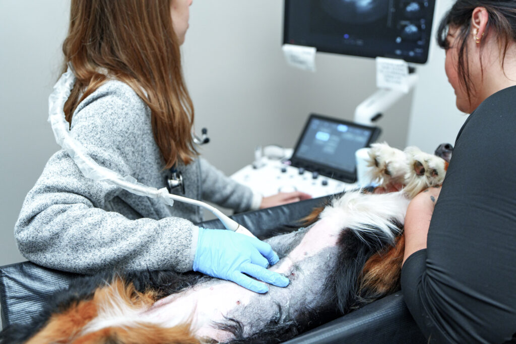
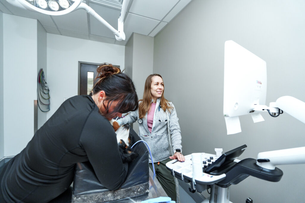
Many dogs and cats can have a routine abdominal ultrasound while awake. They usually rest on a padded table while a technician gently helps them remain in the needed position. A small area of fur is typically clipped because air trapped in the coat interferes with image quality, and ultrasound gel is applied to the skin.
Sedation may be recommended if a pet is very anxious, painful, unable to remain still, or having tissue sampling. The decision is individualized according to temperament, medical condition, and the procedure needed.
How should I prepare my pet?
Follow the hospital’s instructions. For many abdominal ultrasounds, food is withheld beforehand because a full stomach and intestinal gas can limit the view. Water may still be allowed, and a reasonably full bladder can help urinary imaging.
Do not change medications, food, or water access unless the veterinary team specifically directs you to do so. Puppies, kittens, pets with diabetes, and animals with certain illnesses may need modified preparation. Bring a current medication list and any prior images or reports that have not already been transferred.
Ultrasound-guided aspirates and biopsies
Ultrasound can guide a needle into an organ, mass, lymph node, or fluid pocket. A fine-needle aspirate collects cells for cytology; a core biopsy collects tissue for histopathology. These procedures can provide information that imaging alone cannot.
Sampling is not automatically performed during every ultrasound. Before recommending it, the veterinarian considers the target, expected diagnostic value, bleeding risk, current medications, and whether sedation or anesthesia is appropriate. Blood-clotting tests or other preparation may be needed. The benefits, limitations, and potential complications should be reviewed with you first.
How long does an ultrasound take?
The scan often takes approximately 20 to 45 minutes, but timing varies with the body area, findings, patient cooperation, and sampling. The total visit may be longer for record review, examination, preparation, discussion, or sedation recovery.
If your pet is receiving a broader specialty consultation, the ultrasound is one part of the visit rather than the entire appointment.
What happens after the results are available?
The veterinarian interprets the images in the context of the full case. Some findings allow an immediate recommendation; others narrow the possibilities but require laboratory testing, sampling, or monitoring. If an aspirate or biopsy is sent to a laboratory, final results may take additional time.
You should receive an explanation of the important findings, what remains uncertain, and what options come next. Results may lead to medication, diet changes, additional testing, surgery, recheck imaging, or observation. Reports can also be shared with your family veterinarian so that follow-up care is coordinated.
When might an Internal Medicine specialist become involved?
An internist may perform or interpret ultrasound as part of a broader evaluation for chronic digestive disease, endocrine disease, kidney or liver abnormalities, unexplained fluid, weight loss, or other complex concerns. The specialist connects the imaging findings to the history, examination, and laboratory results rather than treating the image as a stand-alone diagnosis.
If symptoms are recurring, several organ systems may be involved, or testing has not explained the problem, read what veterinary internal medicine specialists do and learn about Internal Medicine at COVE.
Frequently asked questions
Is ultrasound safe for dogs and cats?
Diagnostic ultrasound is noninvasive and does not use ionizing radiation. The scan itself is generally painless, although a pet with abdominal pain may be uncomfortable when positioned or gently examined.
Why does my pet need to be shaved?
Sound waves do not travel well through air. Clipping the fur allows the probe and gel to contact the skin, improving image quality and reducing the chance that an important detail is missed.
Can ultrasound prove that a mass is cancer?
Usually not by appearance alone. Ultrasound can identify and characterize a mass, but cytology or biopsy is often needed to determine the cell or tissue type.
Can ultrasound find every swallowed object?
No. Visibility depends on the material, location, gas, and changes caused by the object. Radiographs, repeat imaging, endoscopy, surgery, or other testing may still be necessary.
What if my pet is suddenly very sick?
Ultrasound appointments are not a substitute for emergency evaluation. Trouble breathing, collapse, repeated unproductive retching, inability to urinate, active seizures, toxin exposure, severe pain, or rapid deterioration warrants immediate care. Review COVE’s Emergency Medicine service or call the nearest open hospital.
Talk with COVE about diagnostic imaging
Learn more about Veterinary Radiology and Diagnostic Imaging at COVE. For questions about ultrasound or a specialist evaluation in Central Ohio, contact COVE Pet Care. Your family veterinarian can also send records through COVE’s referral process.
This article is for general educational purposes and does not diagnose your pet or replace an examination by a veterinarian. Preparation, sedation, testing, and treatment recommendations vary by patient.