X-rays and ultrasound are two different ways to look inside a dog or cat. X-rays are especially helpful for bones, joints, lungs, and the overall shape or position of structures. Ultrasound is often better for examining soft-tissue organs, fluid, and movement in real time. A veterinarian may recommend one test or both, depending on the clinical question.
What is the basic difference between an X-ray and an ultrasound?
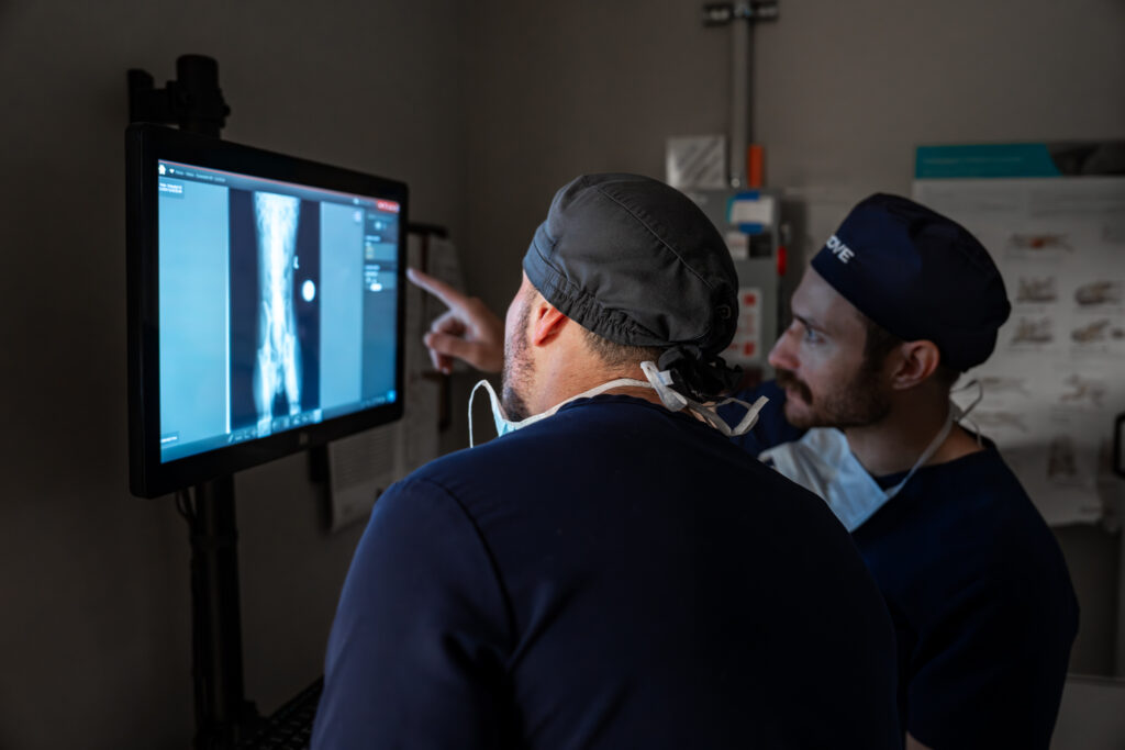
An X-ray, also called a radiograph, uses a small amount of ionizing radiation to create a two-dimensional projection of the body. Air, soft tissue, fluid, and bone absorb X-rays differently, producing a range of black, gray, and white areas.
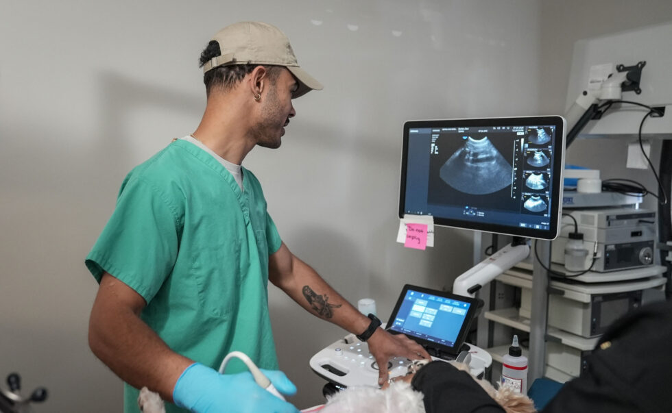
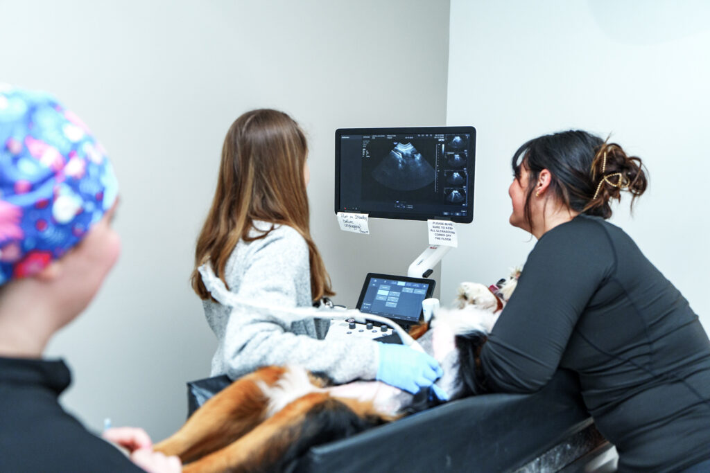
Ultrasound uses high-frequency sound waves rather than radiation. A probe sends sound into the body and receives returning echoes, creating real-time images. The veterinarian can watch organs and some fluids or tissues move during the examination.
What do X-rays show best?
Radiographs provide a broad structural overview. They are commonly used to evaluate:
- Bones, fractures, joint alignment, and some arthritic changes
- The lungs, airways, ribs, diaphragm, and chest cavity
- The size, shape, and position of the heart silhouette and abdominal organs
- Gas patterns within the stomach and intestines
- Some bladder stones and swallowed objects
- Trauma involving the chest, abdomen, spine, or limbs
Because the body is three-dimensional and an X-ray is two-dimensional, the team usually obtains more than one view. Our detailed guide to pet X-rays for dogs and cats explains positioning, sedation, timing, and what owners can expect.
What does ultrasound show best?
Ultrasound is well suited to many internal organs and soft tissues. It can help assess:
- The liver, gallbladder, spleen, kidneys, adrenal glands, and urinary bladder
- The pancreas, lymph nodes, and portions of the stomach and intestines
- Abdominal fluid and some soft-tissue masses
- Organ texture, movement, and blood flow when Doppler is used
- A target for a needle aspirate or biopsy when sampling is appropriate
Gas and bone interfere with sound waves, so ultrasound is generally not the best first study for lungs or the inside of bones. Learn more in our guide to ultrasound for dogs and cats.
Why might a pet need both tests?
X-rays and ultrasound often answer complementary questions. For a vomiting pet, radiographs may show an abnormal intestinal gas pattern, organ enlargement, or dense foreign material. Ultrasound can then provide more detail about the intestinal wall, movement, surrounding fluid, and nearby organs. For trauma, an X-ray may give a useful overview while ultrasound helps the team look for internal fluid.
Needing both tests does not mean the first test failed. Each modality displays anatomy differently, and the combination may help the veterinarian narrow the possibilities or decide what should happen next.
Can either test diagnose cancer by itself?
Not usually. An X-ray or ultrasound may reveal a mass, enlarged organ, abnormal bone, or suspicious tissue pattern, but benign change, inflammation, infection, and cancer can sometimes look similar. Cytology, biopsy, laboratory testing, follow-up imaging, or another procedure may be needed to determine the cause.
Will my pet need sedation?
Many dogs and cats can have radiographs or an abdominal ultrasound while awake. Sedation may be recommended when a pet is painful, very anxious, unable to remain still, or needs precise positioning or sampling. The team considers patient comfort, the information needed, and medication or anesthesia risks before making that recommendation.
How should I prepare my pet?
Preparation depends on the study and your pet’s health. An abdominal ultrasound may require food to be withheld, and a reasonably full bladder can help with some views. Many X-ray studies require no special home preparation, but sedation or another procedure may change the instructions. Always follow the directions from the scheduling or care team, and do not withhold food, water, or medication unless specifically instructed.
How does a veterinarian choose the appropriate test?
The decision starts with the clinical question. The veterinarian considers your pet’s symptoms, examination, medical history, laboratory results, stability, and the body area of concern. X-rays are often an efficient first look at bones, the chest, or an overall abdominal pattern. Ultrasound may be selected when the concern centers on an abdominal organ, soft tissue, fluid, or real-time movement.
Imaging findings are not interpreted in isolation. At COVE, the care team connects them with the rest of the evaluation, and board-certified radiologist review can add specialist expertise when indicated.
When may imaging be urgent?
Immediate evaluation is important when a pet has trouble breathing, collapses, experiences major trauma, repeatedly retches without producing vomit, has a swollen or painful abdomen, cannot urinate, or may have swallowed a dangerous object. Imaging may help after the team assesses and stabilizes the patient. Review COVE’s Emergency Medicine and Critical Care service for urgent care information.
What happens after the results?
Your veterinarian considers the images together with the physical examination, symptoms, and laboratory results. The next step may be monitoring, treatment, repeat imaging, a different imaging method, tissue sampling, surgery, or referral to a specialist. Timing varies with the study and the urgency of the case.
Frequently asked questions
Is ultrasound safer than an X-ray?
Ultrasound does not use ionizing radiation. X-rays do, but veterinary teams use controlled exposures and radiation-safety procedures. The appropriate test is the one most likely to answer the medical question with a reasonable benefit for the patient.
Which test is better for a swallowed object?
It depends on the material, location, and changes it causes. Some objects are visible on radiographs; others are recognized through indirect signs or may be better evaluated with ultrasound. A suspected foreign body obstruction requires prompt veterinary guidance.
Can ultrasound replace X-rays?
No single imaging method is best for every problem. Ultrasound is limited by gas and bone, while X-rays provide less soft-tissue detail. They frequently complement one another.
Can owners choose which test their pet receives?
Your observations and questions are important, but the veterinarian recommends imaging based on the clinical concern and the information needed. Ask what each proposed test may show, what its limits are, and how the result could change the plan.
Talk with COVE about diagnostic imaging
Learn about Veterinary Radiology and Diagnostic Imaging at COVE. For a current COVE patient, specialty consultation, outpatient ultrasound, or referral questions in Central Ohio, contact COVE Pet Care.
This article is educational and is not a substitute for an examination or diagnosis by a veterinarian. If your pet is ill, injured, or worsening, contact a veterinary professional promptly.