Osteochondritis dissecans (OCD) is a joint condition in which abnormal cartilage development leads to a weakened, cracked, or lifted cartilage flap. The lesion can cause inflammation, pain, lameness, and progressive arthritis. OCD is part of a broader developmental process called osteochondrosis.
The condition is most often recognized in growing medium, large, and giant-breed dogs. The shoulder is commonly affected, but lesions can also occur in the elbow, knee, and hock. Diagnosis and treatment depend on the joint, lesion size and location, symptoms, age, arthritis, and other orthopedic findings.
Osteochondrosis Versus Osteochondritis Dissecans
Osteochondrosis is a disturbance in normal cartilage-to-bone development near a joint. The cartilage becomes abnormally thick or weak. When cracks form and a cartilage flap separates from the underlying bone, the condition is called osteochondritis dissecans, or OCD.
A flap may remain attached, become unstable, or break free within the joint. Damaged cartilage and exposed underlying bone trigger inflammation and can lead to osteoarthritis.
Why OCD Develops
The exact cause is not always known. Rapid growth, genetics, nutrition, joint loading, minor trauma, and other biological factors may contribute. Large and giant breeds are represented more often, but breed and size alone do not establish a diagnosis.
OCD is developmental rather than simply an injury caused by normal exercise. Owners should not assume they caused the condition with one walk, jump, or play session. The opposite joint may also need evaluation because lesions can occur on both sides.
Joints Commonly Affected
Shoulder
Shoulder OCD often causes front-leg lameness in a young, growing dog. The limp may worsen with exercise and improve with rest. Both shoulders can be affected even when one side appears more painful.
Elbow
Elbow osteochondrosis can occur with other developmental elbow conditions. Dogs may show front-leg lameness, stiffness, reduced range of motion, or an altered stance. CT is often helpful because the elbow is complex and overlapping bones can limit radiographic detail.
Knee
OCD of the knee, or stifle, can cause back-leg lameness, joint swelling, pain, and stiffness. CCL disease, patellar luxation, meniscal injury, and other knee conditions can produce similar signs, so a full orthopedic assessment is important.
Hock
Hock lesions may cause swelling, stiffness, and back-leg lameness. Joint location and lesion shape can affect treatment and long-term expectations.
Symptoms of OCD in Dogs
- Intermittent or persistent limping
- A limp that worsens after exercise
- Stiffness after rest
- Joint swelling or fluid accumulation
- Pain with joint movement
- Reduced range of motion
- Reluctance to run, jump, climb stairs, or play
- Muscle loss when the condition is chronic
These signs can also come from CCL disease, patellar luxation, arthritis, fracture, muscle injury, or another developmental joint disorder. Review the guide to common causes of dog limping for broader warning signs and emergency guidance.
How OCD Is Diagnosed
History and Orthopedic Examination
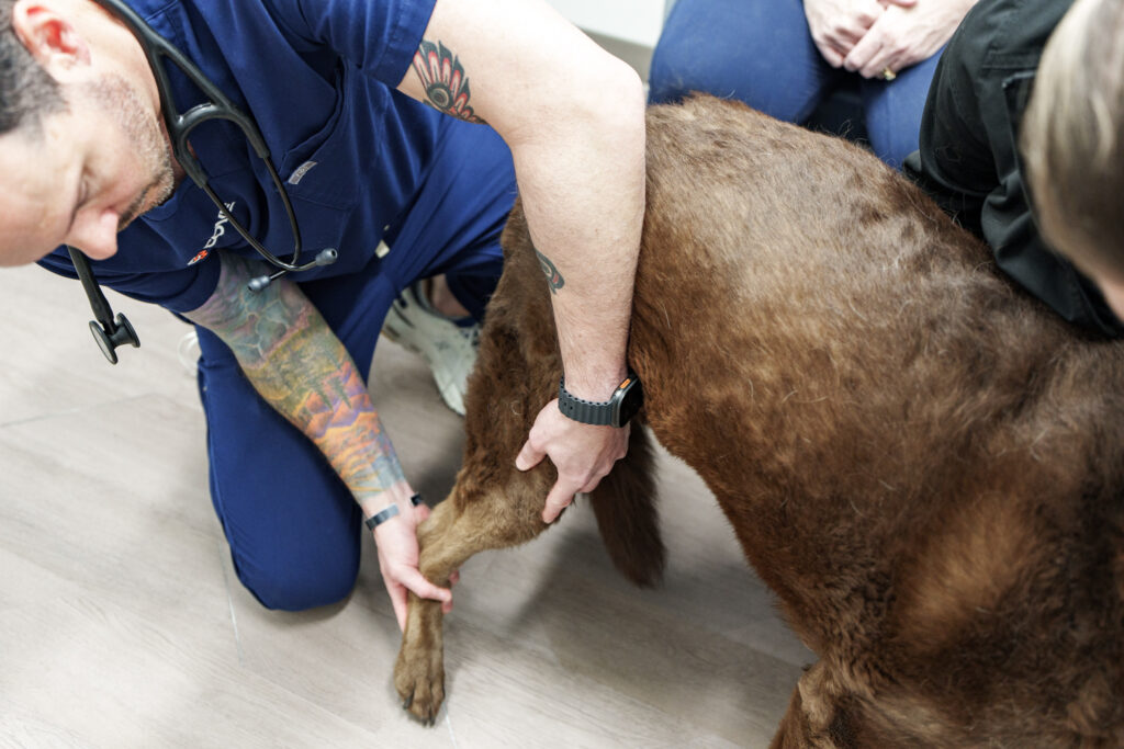
The veterinarian considers age, breed, growth history, when the limp started, whether it changes with activity, and whether both sides may be involved. The orthopedic examination assesses gait, joint swelling, pain, range of motion, muscle mass, and other possible sources of lameness.
Radiographs
Radiographs may show flattening, a defect in the bone beneath cartilage, a fragment, joint swelling, or arthritis. Specific positioning is important, and sedation may be recommended for accurate images and patient comfort. Both sides may be imaged for comparison.
CT, Arthroscopy, and Other Imaging
CT provides detailed cross-sectional images and can help define lesion size, location, fragments, and bone changes, especially in complex joints. Arthroscopy allows the surgeon to look directly inside the joint and may combine diagnosis with treatment. Additional imaging depends on the joint and differential diagnoses.
Does Every OCD Lesion Need Surgery?
No. Treatment decisions consider symptoms, lesion stability, location, size, patient age, joint affected, arthritis, and evidence of healing. Small, early lesions in selected young dogs may be managed with activity restriction and a veterinarian-directed plan.
Persistent lameness, an unstable flap, a free fragment, a large defect, or a lesion unlikely to heal may make surgery more appropriate. Online information cannot determine whether an individual lesion should be monitored or treated surgically.
Surgical Treatment for OCD
Removing Unstable Cartilage
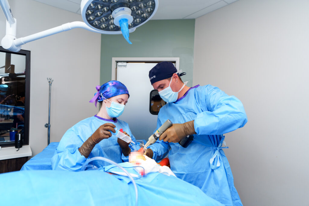
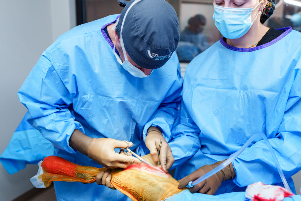
The surgeon may remove a loose flap or fragment that is causing inflammation and mechanical irritation. This can be performed through arthroscopy or an open approach depending on the joint, lesion, equipment, and surgeon’s plan.
Stimulating Healing
The abnormal bed beneath the cartilage may be debrided or curetted to encourage bleeding and formation of repair tissue. The new tissue is not identical to normal joint cartilage, and arthritis may still develop.
Joint Resurfacing
Selected large or difficult lesions may be treated with an osteochondral graft or another resurfacing technique intended to restore a more durable joint surface. These procedures are not needed for every OCD case and require detailed assessment and planning.
Recovery After OCD Surgery
Recovery depends on the joint, lesion, and procedure. The plan may include confinement, leash-only activity, incision protection, prescribed medication, rehabilitation, rechecks, and follow-up imaging. Running, jumping, stairs, and rough play are restricted until the surgical team allows progression.
Rehabilitation may help restore joint motion, muscle strength, balance, and controlled limb use. Exercises should be selected for the specific joint and healing stage. The dog’s discharge instructions always override a general online timeline.
Arthritis and Long-Term Expectations
OCD damages a joint surface and can lead to osteoarthritis. Surgery may improve comfort and function by removing unstable tissue and treating the defect, but it cannot guarantee a normal joint or eliminate future arthritis.
Outcome varies with the affected joint, lesion size and location, duration, arthritis, age, and treatment. Long-term care may include healthy weight management, regular appropriate exercise, rehabilitation, and a veterinarian-directed arthritis plan.
What Owners Should Ask
- Which joint and surface are affected?
- Is the lesion stable, attached, or free within the joint?
- Should the opposite joint be imaged?
- Would CT or arthroscopy change the plan?
- Is monitoring reasonable, or is surgery recommended?
- What procedure is planned and why?
- What activity restrictions and rehabilitation are expected?
- How might arthritis affect long-term function?
OCD Evaluation and Surgery at COVE
COVE Pet Care evaluates joint pain and developmental orthopedic conditions for dogs in Columbus, Clintonville, Delaware, and across Central Ohio. The orthopedic team examines the full limb, reviews imaging, and explains whether monitoring, rehabilitation, arthroscopy, or another surgical approach may be appropriate.
Explore orthopedic surgery at COVE or request a consultation. If a dog has sudden severe lameness after trauma, cannot stand, or has another emergency sign, seek prompt veterinary care.