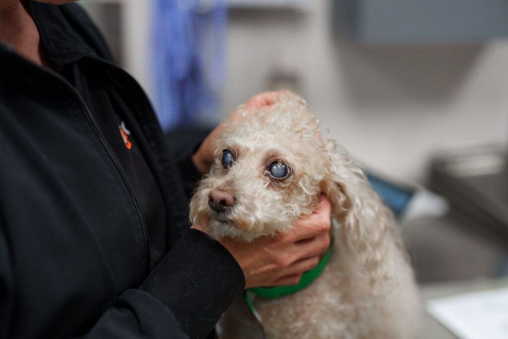
A dog’s cloudy eye can have many causes. It may be a normal lens change, a cataract, corneal swelling, or high eye pressure. Swelling inside the eye can also cause haze. These problems may look alike from across the room. They need very different care.
Call a vet soon if cloudiness starts fast or comes with redness or squinting. A wide pupil, a large eye, vomiting, low energy, or a change in sight are also urgent. Do not use human drops or old pet medicine. An eye exam is the only safe way to find where the cloudiness starts.
The short answer
Cloudiness may sit on the eye’s clear front, inside the lens, or deeper in the eye. A vet may use a stain, a bright light, and an eye-pressure test. A slow blue haze in both eyes may be normal aging. A sudden, red, or painful haze needs quick care.
What you can watch at home
Check both eyes in soft light. Note if the haze is on the clear front or seems deep behind the pupil. Do not shine a harsh light in a sore eye. Watch your dog walk through a known room and then a dim room. Look for fear at steps, doors, and curbs. Write down when the change began and if it is getting worse. A photo may help show change over time, but it cannot test pain, pressure, or sight. Call the vet if the eye turns red, the pupil stays wide, or your dog rubs the face.
Keep your dog on a leash near roads and steps until sight is checked. Leave food, water, and beds in the same place. These small steps can lower stress while you arrange care.
Trust a new change, even if your dog still acts well. Dogs can hide pain and use smell to cope with poor sight.
Where cloudiness can develop
Owners often describe any pale, blue, gray, or white appearance as a “cloudy eye.” The change may actually be located in one of several structures:
- Cornea: the transparent outer window at the front of the eye
- Anterior chamber: the fluid-filled space behind the cornea
- Lens: the normally clear focusing structure behind the pupil
- Vitreous, retina, or deeper tissues: structures toward the back of the eye
Good light may hint at the location. A home check cannot replace a vet exam. Bright light, a close view, and an eye-pressure test help find the true cause.
Cataracts
A cataract is an opacity within the lens. It may affect a small portion of the lens or progress until much of the pupil looks white or opaque. Cataracts can reduce vision and may trigger inflammation inside the eye. Causes include inherited disease, diabetes, trauma, inflammation, retinal disease, and aging.
Dogs with cataracts may fear stairs, struggle in dim light, or bump into things. Not every cataract needs surgery. Not every eye can have surgery. An eye specialist checks the retina, the eye, and the dog’s health. Home care also matters. COVE’s guide explains cataract signs, surgery tests, lens removal, and recovery.
Nuclear sclerosis
Nuclear sclerosis, also called lenticular sclerosis, is a common age-related change. New lens fibers continue to form throughout life, compressing older fibers toward the center. Both pupils may develop a transparent blue-gray haze, often gradually and at a similar pace.
Unlike a dense cataract, nuclear sclerosis usually does not block vision, although close focusing may become less precise. The distinction can be difficult for owners to make because both changes sit behind the pupil. Cornell University’s canine cataract information describes how a veterinary exam separates cataracts from normal lens aging.
Corneal disease
A cloudy or blue surface may mean that fluid has built up in the cornea. This is called corneal edema. It can occur with an ulcer, injury, swelling, a loose lens, or glaucoma. Fat, calcium, pigment, cells, or scar tissue can also make the cornea less clear.
A corneal ulcer frequently causes squinting, tearing, redness, and rubbing. Some defects remain shallow, but infected or “melting” ulcers can deepen rapidly. Learn how vets use fluorescein stain and other tests in the guide to corneal ulcers in dogs and cats.
Glaucoma
Glaucoma is abnormally high pressure inside the eye. Pressure can cause diffuse blue-gray corneal cloudiness, redness, a dilated pupil, pain, and vision loss. Over time the eye may enlarge, but an acute attack can begin before enlargement is obvious. Some dogs appear quiet, nauseated, or unwilling to eat rather than showing unmistakable eye pain.
Suspected glaucoma is an emergency because pressure can damage the retina and optic nerve. If cloudiness is sudden or painful, seek care immediately. Read more about glaucoma warning signs, pressure testing, and treatment goals.
Uveitis and inflammation inside the eye
Uveitis is swelling in the colored tissue inside the eye. Protein and cells can leak into eye fluid and cause haze. Other signs include redness, pain, a small or odd pupil, and poor sight. Causes include injury, lens disease, infection, immune disease, cancer, and illness elsewhere in the body.
Because the eye change may be connected to systemic health, the vet may recommend bloodwork, imaging, infectious-disease testing, or other exam.
How quickly does a cloudy eye need care?
Seek same-day or emergency exam if you observe:
- Cloudiness that appeared over hours or a day
- Squinting, rubbing, or holding the eye closed
- Marked redness or swelling
- A pupil that is enlarged, very small, irregular, or different from the other pupil
- An enlarged, bulging, injured, or displaced eye
- Sudden bumping, hesitation, disorientation, or other vision change
- Vomiting, severe lethargy, or loss of appetite with an abnormal eye
A slow, painless haze in both eyes may be less urgent, but it still deserves an exam. Monitoring a photograph over months cannot determine whether pressure, inflammation, or retinal function is changing.
Tests that distinguish the causes
A vet may check sight, pupils, tears, the cornea, the lens, pressure, and the retina. A bright stain shows many ulcers. A pressure test helps separate glaucoma from problems that lower eye pressure. A strong light helps locate the cloudy area.
When a dense cataract blocks the view into the eye, ocular ultrasound can assess the retina and other deeper structures. Electroretinography may be recommended to measure retinal function before cataract surgery. Additional testing depends on the eye findings and the dog’s general health.
What treatment may involve
Care targets the cause, not the cloudy look. An ulcer may need eye protection, pain relief, and medicine for germs. Glaucoma needs fast pressure control. Uveitis needs care for swelling and its cause. Cataracts may be watched or checked for surgery. Normal lens aging needs no care.
Some medicines that are useful for one condition are unsafe for another. In particular, a steroid-containing drop can delay corneal healing or worsen certain infections. Never start or restart an eye medicine without veterinary direction and a current exam.
Getting the right level of care
Primary vets diagnose and treat many eye problems. Referral may help when cloudiness is severe, the diagnosis is uncertain, vision is at risk, advanced testing is needed, or surgery may be considered. Learn when a pet should see a veterinary ophthalmologist.
If your dog’s eyes look cloudy, contact a veterinary team rather than relying on appearance alone. For specialist capabilities, team information, locations, and verified contact details, visit COVE’s Veterinary Ophthalmology service page. Online information can help you recognize warning signs, but it cannot identify the cause or predict your dog’s vision.