Glaucoma causes pressure inside the eye to rise too high. This harms the retina and optic nerve. It is painful and can cause lasting vision loss. Do not assume that a red, cloudy, large, or blind eye has simple irritation.
Possible glaucoma is an emergency. Get care right away for sudden haze, a wide pupil, severe redness, or hard squinting. A large eye or fast vision change is also urgent. Vomiting or low energy can be a sign of eye pain. Do not use human drops or old medicine. Only an exam can measure eye pressure and sight.
The short answer
Glaucoma is high pressure inside the eye. Fast care may ease pain and help save sight. A vet uses a small tool to check the pressure. Medicine may lower it. Some pets need surgery. If sight is already gone and pain will not stop, the main goal becomes long-term comfort.
What to do when glaucoma may be present
Call a vet or emergency clinic at once. Say that the eye is red, cloudy, large, or painful. Tell them if the pupil stays wide or sight has changed. Share signs such as vomiting, hiding, or loss of food drive. Do not press on the eye. Keep your pet in a calm, dim room while you get ready to leave. Bring all eye drops and pills. If your pet has had glaucoma before, bring the past pressure notes if they are easy to reach.
Do not wait for the eye to look better on its own. A pet may stop squinting even while pressure stays high. Only a pressure test can show if the eye is safe.
Keep the trip calm and quiet. Use a carrier for a cat and a leash for a dog. Do not let your pet rub the face. Ask the clinic where to go if the first site cannot check eye pressure. Fast care is more important than waiting for a past vet or a set visit time.
Call now. Do not wait.
How eye pressure becomes elevated
Clear fluid called aqueous humor is continually produced inside the front of the eye. It circulates through the pupil and leaves through a drainage angle where the iris and cornea meet. Glaucoma develops when drainage is impaired and fluid pressure rises.
The problem is not just “too much fluid.” High pressure harms the retina and optic nerve. These tissues send visual signals to the brain. The amount of harm depends on how high the pressure is and how long it stays high.
Warning signs of glaucoma
- A red or bloodshot eye
- Blue-gray corneal cloudiness
- A pupil that is dilated or responds poorly to light
- Squinting, rubbing, light sensitivity, or resistance to head touch
- Sudden bumping, hesitation, disorientation, or vision loss
- An eye that looks larger or firmer than the other
- Lethargy, hiding, poor appetite, nausea, or vomiting
Long-term glaucoma may be harder to spot. The eye may slowly grow or stay hazy. Sight may be gone before the change looks clear. Cats may only act quiet or avoid touch. Any lasting difference between the eyes needs an exam.
Primary and secondary glaucoma
Primary glaucoma
Primary glaucoma starts with an inherited or early problem in the eye’s drain. Some dog breeds face more risk. One eye may be affected first, but the other eye can be at risk. The eye specialist may advise medicine and checks for that eye. No plan can promise that glaucoma will not start.
Secondary glaucoma
Secondary glaucoma occurs when another eye disease blocks fluid drainage. Causes include inflammation inside the eye, lens displacement, advanced cataract-related change, bleeding, trauma, scar tissue, retinal detachment, and tumors. In these cases, treatment must address both the pressure and the underlying disease when possible.
In cats, glaucoma is often secondary to chronic inflammation or other ocular disease. The affected eye may enlarge gradually and signs may be missed until vision has been lost. The Merck Veterinary Manual overview of glaucoma in small animals explains the major primary and secondary forms.
How glaucoma is diagnosed
Tonometry is a quick eye-pressure test. The number is only one part of the exam. The vet also considers the cornea, swelling, species, test method, and time of day. One pressure result does not prove the full cause.
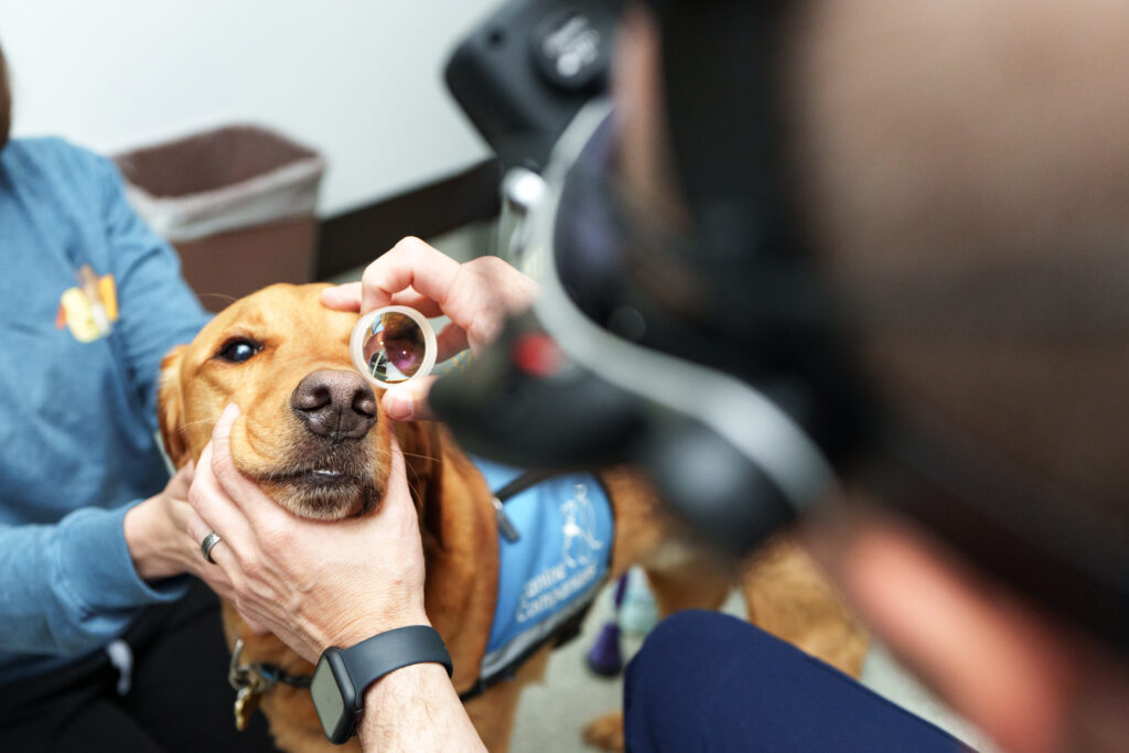
The vet also checks sight, pupils, the cornea, lens, retina, optic nerve, and pain. A special lens can look at the eye’s drain. This may help show risk in the other eye. Ultrasound can help when haze blocks the view. It may also show a mass, loose lens, or detached retina.
Bloodwork or other testing may be recommended when inflammation, infection, cancer, or systemic illness could be contributing. COVE’s article on veterinary eye-exam tests explains common tests and how owners can prepare.
Emergency treatment goals
If useful sight remains, the first goal is to lower pressure fast. This may protect the retina and optic nerve. Care may include several eye drops and medicine by mouth or injection. The plan must fit the pet’s heart, kidneys, and health. Pain and nausea also need care.
The response is measured with repeat pressure checks. An eye that looks less red is not necessarily at a safe pressure. If medical treatment cannot control pressure or if the anatomy makes recurrence likely, the ophthalmologist may discuss a procedure or surgery.
Procedures for an eye with vision
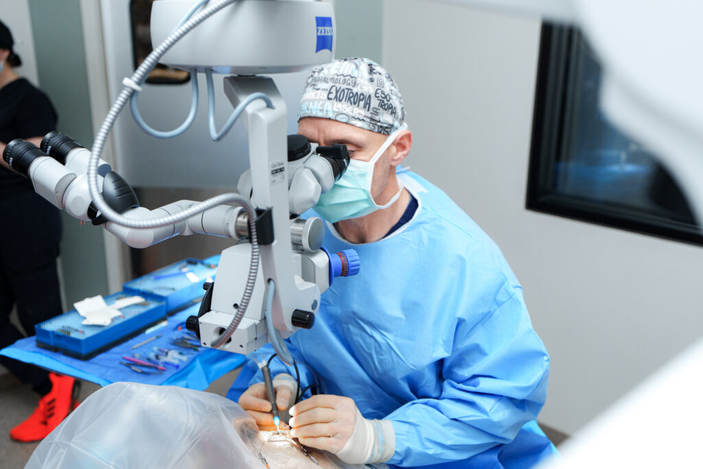
Surgical choices vary with the type of glaucoma, remaining vision, eye structure, and underlying disease. Procedures may aim to reduce fluid production, improve outflow, or both. Examples include laser treatment of the fluid-producing ciliary body and placement of a drainage implant. These procedures can extend pressure control in selected patients, but glaucoma can recur and continued monitoring is essential.
Cataract, lens displacement, inflammation, or other disease may change the plan. The ophthalmologist will discuss realistic benefits, aftercare, possible complications, and the likelihood that additional medicine or procedures will be needed.
Keeping a blind, painful eye comfortable
If sight cannot return and pressure still hurts, the goal changes to comfort. Medicine may help for a time. Some pets need the painful eye removed. This choice can feel hard, but pets often adjust very well once the pain is gone.
Cosmetic considerations, pathology testing, recovery, and what to expect at home should be discussed before surgery. Choosing comfort is not a failure of treatment; in a blind eye with uncontrolled glaucoma, it can be the most humane and reliable goal.
What about the other eye?
With primary glaucoma in dogs, the second eye may be vulnerable even when it looks normal. The ophthalmologist may recommend gonioscopy, baseline pressure measurements, preventive medicine, and scheduled rechecks. Owners should learn the signs of an acute attack and know where to seek urgent care.
Secondary glaucoma does not always carry the same risk to the other eye, but the underlying cause matters. Bilateral inflammation, inherited lens disease, or systemic illness can affect both eyes. An individualized diagnosis is more useful than a general prediction.
When specialist care helps
Glaucoma often requires urgent pressure control, advanced exam, long-term monitoring, and discussion of medical and surgical options. Learn more about what a board-certified veterinary ophthalmologist does. If the eye is suddenly painful or vision has changed, follow the guidance in the pet eye emergency guide and seek care now. COVE also explains other causes of sudden blindness in dogs.
For COVE’s eye-care capabilities, team, Central Ohio locations, and verified contact details, visit the COVE Ophthalmology service page. Glaucoma cannot be diagnosed or safely managed through photographs or online advice; the eye’s pressure and health must be assessed in person.