A cloudy eye does not always mean a dog has a cataract. Still, cataracts can grow and reduce sight. They can also cause painful swelling inside the eye. A veterinary exam is the only safe way to find the cause. COVE’s guide compares other common causes of cloudy eyes in dogs.
This guide compares cataracts with normal lens aging. It also explains surgery tests and recovery. Get prompt care for sudden cloudiness, severe redness, squinting, a wide pupil, or sudden vision loss. Do not use human eye drops or old medicine. Use only medicine prescribed for your dog.
The short answer
A cataract is a cloudy spot inside the lens. It can make sight dim or block it fully. A vet can tell a cataract from normal lens aging. Some dogs do well with close eye care and medicine. Others may be able to have surgery. The best plan depends on the eye, the retina, the dog’s health, and the family’s ability to give care at home.
Watch how your dog moves in dim light and on stairs. Note if one eye changes faster than the other. Share these clues with your vet. They can help show how much the change affects daily life.
What is a cataract?
The lens is a clear structure behind the iris and pupil. It focuses light on the retina at the back of the eye. A cataract is a cloudy area within the lens. A small cataract may have little effect on sight. A large cataract can block light and cause partial or full vision loss.
Cataracts are more than a change in looks. Lens proteins can cause painful swelling called lens-induced uveitis. Old cataracts may also lead to glaucoma, a loose lens, or retinal detachment. An early eye exam helps the specialist watch for these problems. It also gives the family more time to discuss surgery.
Cataracts versus nuclear sclerosis
Many older dogs develop a blue-gray haze called nuclear sclerosis. It is also known as lenticular sclerosis. This normal change happens as lens fibers become packed together with age. Most dogs keep useful sight, though close focus may change.
A cataract is a true cloudy spot that can block light from the retina. Both problems may look pale or hazy behind the pupil. A vet can tell them apart with bright light and a close view of the lens. Do not assume that cloudiness is harmless. Call a vet if your dog bumps into things, fears stairs, or seems uncomfortable.
Cornell University’s canine cataract overview also explains the difference between cataracts and lenticular sclerosis.
What causes cataracts in dogs?
Many cataracts are inherited. The age when they appear can vary by breed and family line. Other causes include diabetes, eye swelling, injury, poor nutrition, retinal disease, and aging.
Diabetes and rapid cataract formation
Many dogs with diabetes develop cataracts, sometimes very fast. Extra sugar in the lens changes into sorbitol. This substance pulls water into the lens and harms its fibers. Sight may worsen over days or weeks. Diabetic control and overall health affect anesthesia. Eye health affects whether surgery is likely to restore useful sight.
A dog does not need to become fully blind before an exam. Swelling and other changes can make surgery harder as the cataract grows. An early talk with an eye specialist can help.
Symptoms of cataracts and related complications
Possible signs include:
- A white, gray, or opaque appearance behind the pupil
- Difficulty seeing in dim light or unfamiliar spaces
- Hesitation at stairs, curbs, or doorways
- Bumping into furniture or people
- Startling more easily when approached
- Redness, squinting, tearing, or sensitivity to light
Dogs often adapt well to slow vision loss. They use hearing, smell, memory, and whiskers. This can make the change hard to notice. Pain is never a normal part of cataracts. Redness or squinting may point to swelling, glaucoma, corneal disease, or another urgent problem.
How is a dog checked for cataract surgery?
Surgery may be considered when a cataract harms useful sight. The eye, the dog, and the family must also be ready for surgery and home care. Testing often includes a full eye exam, a tear test, a corneal check, and an eye-pressure test. The specialist also looks for swelling, glaucoma, retinal disease, and other problems that may affect the outlook. Read more about veterinary eye tests and diagnostics.
Blood work, a urine test, and a physical exam help check overall health and anesthesia risk. Some dogs need more tests. A diabetic dog may need recent blood-sugar records and an infection check. The plan is made for each dog.
Ocular ultrasound
A dense cataract can block the doctor’s view of the retina. An eye ultrasound uses sound waves to look behind the cataract. It can show a detached retina, a loose lens, or changes in the gel inside the eye.
Electroretinography
An electroretinogram, or ERG, checks how the retina responds to light. A healthy retina is needed for sight after the cloudy lens is removed. If the retina works poorly, surgery may not restore useful sight. This can be true even when the surgery itself goes as planned.
The Ohio State Veterinary Medical Center’s cataract surgery guide describes ophthalmic exam, ERG, ocular ultrasound, and general-health testing used when evaluating candidates.
How cataract surgery is performed
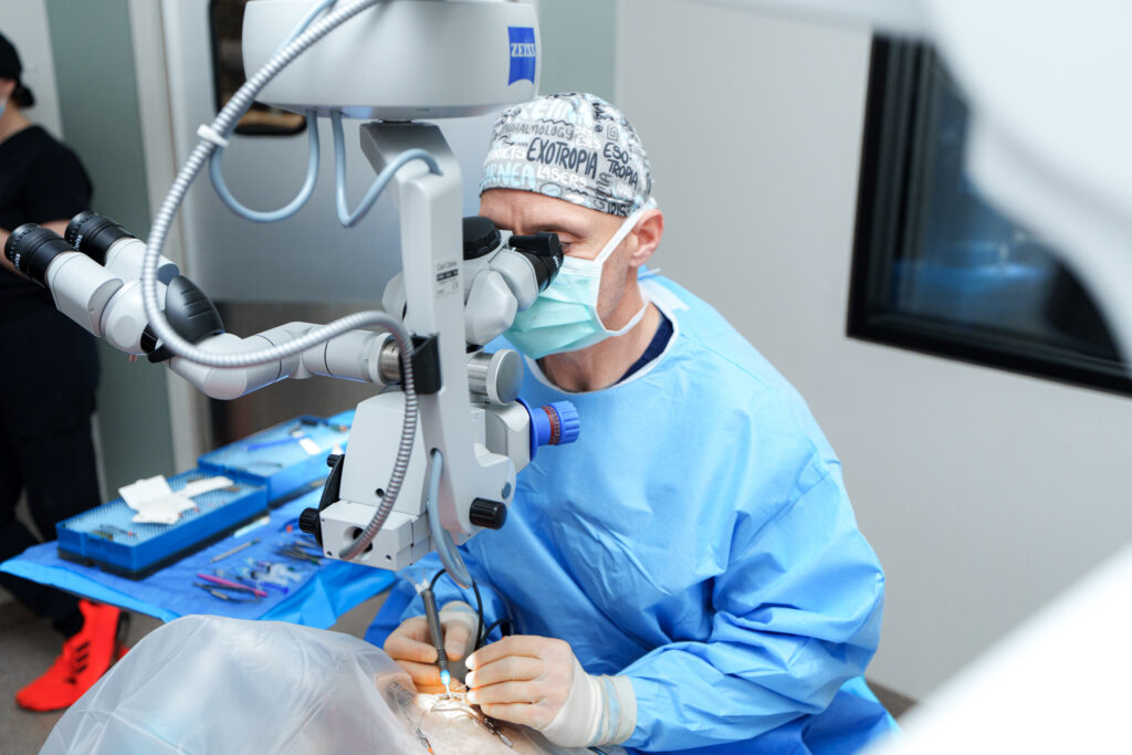
Cataract surgery is done under general anesthesia. The surgeon uses a microscope and makes a small cut in the cornea. The surgeon then opens the thin bag around the lens. A tool uses sound energy to break up the cloudy lens. Fluid and lens pieces are then removed.
If the lens bag is healthy, the surgeon may place a folded plastic lens inside it. The new lens helps the eye focus. The small cut is closed with very fine stitches. Some dogs have both eyes treated at one time. Others have one eye treated based on health and family goals.
Recovery and follow-up after cataract surgery
Recovery takes careful work at home. Most dogs need several eye medicines and may also need pills. An Elizabethan collar protects the eye. Activity must be limited, and early rechecks are common. The medicine schedule can be demanding during the first weeks.
At rechecks, the specialist measures eye pressure and checks healing. Medicine may be changed as the eye heals. Some dogs need long-term drops or later checkups. Before surgery, make sure each caregiver can follow the full schedule.
Possible risks and realistic expectations
No surgery can promise sight. Risks include swelling, glaucoma, infection, a corneal ulcer, bleeding, a detached retina, and vision loss. Some problems can be treated when found early. Others may be serious even with good care.
The outlook depends on retinal function, eye health, overall health, and cataract stage. The cause and response to swelling also matter. Home medicine and rechecks are a major part of success. The goal is useful, comfortable sight, not perfect sight. Dogs that cannot have surgery still need care to control pain and swelling.
Apricot’s story: cataract surgery after diabetes-related blindness
Case study: Apricot developed diabetes while her family was away helping a relative after surgery. When the family came home, Apricot was blind. Her diabetes had caused cataracts to grow very fast.
Eric J. Miller, DVM, MS, DACVO, checked Apricot’s eyes and retina. He needed to know if surgery was a good option. After testing, Dr. Miller removed the cataract with phacoemulsification. He kept the lens bag and placed a folded plastic lens inside it. Apricot’s story shows how fast diabetic cataracts can change sight. It also shows why each dog needs a full specialist exam before surgery.
One dog’s story cannot predict another dog’s result. Cataract type, retinal health, swelling, anesthesia risk, and home care can all differ.
When should you request an ophthalmology exam?
Ask for an eye exam when you first notice lasting lens cloudiness or a change in sight. Early care is key after a diabetes diagnosis. Get prompt help if cloudiness starts fast. Redness, squinting, a large eye, a wide pupil, vomiting, low energy, or clear pain also need quick care.
Learn when a pet should see an eye specialist. You can also review COVE’s eye services, team, Central Ohio locations, and contact details. An online article cannot tell if your dog has a cataract or can have surgery. That requires an exam and a personal talk with a vet.