A veterinary eye exam answers a few key questions. Does the eye hurt? Can the pet see? Which part is not normal? Is eye pressure safe? Is the cornea whole? Does the problem affect one eye, both eyes, or the rest of the body? The team chooses tests that give clear answers with as little stress as possible.
Most basic tests are quick and can be done while a pet is awake. A scan or procedure may need light sleep or anesthesia. This depends on the pet and the test. Get urgent care for sudden blindness, severe pain, injury, a bulging eye, or fast cloudiness. Do not wait for a routine visit.
The short answer
Many eye tests take only a few minutes. A paper strip checks tears. A bright dye shows corneal wounds. A small tool checks eye pressure. Lights and lenses help the doctor see the lens and retina. Some pets also need an ultrasound or ERG. The test plan depends on the signs and exam.
Before the appointment
Bring a current medicine list. Include the name and timing of each drop, pill, and supplement. Ask other clinics to send exam notes, lab results, scans, pressure readings, and surgery reports. Photos can show when a change began, but they do not replace an exam.
Do not stop prescription medicine unless the veterinary team tells you to. Ask before giving scheduled eye drops on the day of the visit, because certain medicines or stains can influence testing. Bring a favorite reward if dietary restrictions allow it, and use a secure leash or carrier.
History and observation
The team will ask when signs began and how they have changed. They will ask which eye is affected and if sight seems different. Tell them about an injury or chemical contact. Diabetes, high blood pressure, immune disease, cancer, and infection can affect the eyes. Breed, age, travel, and family history may matter too.
The exam starts before any tool touches the eye. The vet looks at face shape, eye position, lids, blink, mucus, and pupils. Head position and movement through the room also give clues.
Vision responses and reflexes
Simple checks help show where vision may be lost. The vet may see if a pet tracks a soft cotton ball or moves around an object. A hand test checks for a blink without touch or a puff of air. Pupil checks show how the eye and brain respond to light.
A reflex is not the same as full sight. Age, fear, face-nerve disease, and brain disease can change the result. The vet reads all of the checks as one set.
Schirmer tear test
The Schirmer test measures the watery part of tears. A thin paper strip sits inside the lower lid for one minute. The vet records how far the tears travel. That result is compared with the eye surface and the pet’s signs.
Low tear flow can support a dry eye diagnosis. Tear results can vary. Poor tear quality or weak blinking can still harm the surface when one test is not very low. Read about dry eye signs, tests, and long-term care.
Fluorescein stain
Fluorescein is a bright dye that sticks to an open corneal wound. It does not stick to a healthy outer layer. The dye shows many ulcers and their shape. It can also help check if tears drain into the nose.
A positive stain changes which medicines are safe. Steroid drops can slow healing or worsen some germs when an ulcer is present. A very deep wound needs care when the stain is read. Its thin center may not hold dye. The guide to corneal ulcers in dogs and cats explains why depth matters.
Tonometry
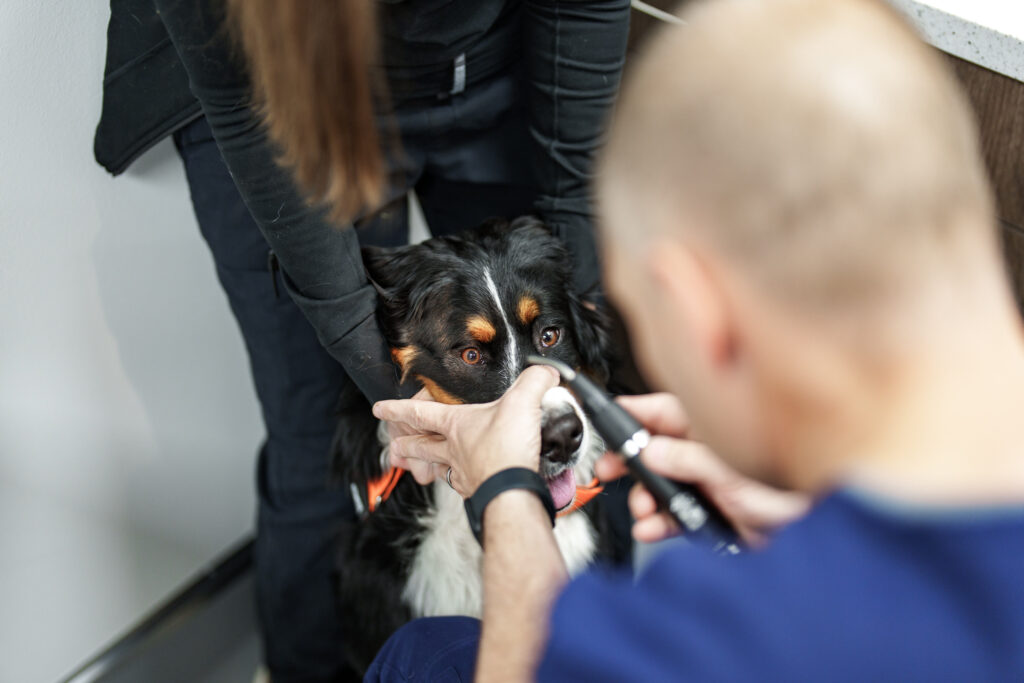
Tonometry measures pressure inside the eye. After numbing drops when needed, a small instrument gently touches or briefly rebounds from the cornea. The test takes seconds. Pressure that is too high raises concern for glaucoma; pressure that is low can occur with uveitis or severe inflammation.
The vet reads the number with the rest of the exam. Test method, corneal health, lid force, species, and time of day can change it. A repeat check can confirm the result and track care. Possible glaucoma needs an urgent pressure check.
Slit-lamp biomicroscopy
A slit lamp makes a narrow beam and a close view of the eye. It shows the lids, cornea, front chamber, iris, and lens. The doctor changes the light angle and width. This can show the depth and place of a spot that only looks “cloudy” at home.
This exam can show corneal layers, cells in eye fluid, iris change, odd lashes, cataracts, and a loose lens. The pet stays awake and is held with care.
Dilated retinal exam
Drops make the pupil wide so the vet can see the lens, eye gel, retina, blood vessels, and optic nerve. A special light gives a broad view of the back of the eye. The drops can last for hours. Bright light may bother the pet for a short time.
The exam may show retinal wear, a detached retina, blood, swelling, germs, pressure harm, or an optic-nerve change. A dense cataract or corneal haze can block the view. In that case, ultrasound or an ERG may help.
Gonioscopy
Gonioscopy uses a special lens to view the eye’s drain. The drain cannot be seen in a normal front view because light bends at the cornea. This test can show risk for primary glaucoma in some dogs. It may also guide care.
The test shows risk. It cannot say if or when glaucoma will start. The vet also looks at breed, family line, eye pressure, and both eyes.
Ocular ultrasound
Ultrasound uses sound waves to make a view inside the eye. It helps when a cataract, blood, swelling, or corneal disease blocks the retina. The scan may show a detached retina, loose lens, changed eye gel, mass, or odd eye shape.
Numbing drops and a soft touch are often enough. A pet may need light sleep if motion would make the scan unsafe. Ultrasound shows shape. It does not prove that the retina can sense light.
Electroretinography
An electroretinogram, or ERG, checks how the retina responds to flashes of light. It may be used before cataract surgery when the retina cannot be seen. It can also help with sudden blindness when the retina first looks normal. This test is key for confirming SARDS in dogs.
The room is kept dark before the test. A special lens and set light flashes are used. Some pets need light sleep or brief anesthesia. An ERG checks the retina, not the whole path from the eye to the brain.
Additional testing
Eye findings may lead to a wider health check. The vet may test blood, urine, blood pressure, or germs. Some pets need scans, cell samples, culture, or a tissue sample. CT, MRI, or nerve tests may also help. The choices depend on the likely cause.
Ohio State’s animal eye-care guide lists tests and care for cataracts, glaucoma, corneal disease, retinal disease, and other eye problems.
What happens after testing?
The eye specialist explains the cause or most likely causes. The team will tell you if sight or comfort is at risk. The plan may include medicine, surgery, more tests, or close checks. Ask which changes mean you should call. Also ask when pressure, stain, or another test must be checked again.
Learn what to expect from an eye specialist and when referral helps. Visit the COVE Ophthalmology service page for the team, tests, care, Central Ohio sites, and contact details. This guide can help you plan for a visit. Your vet must choose and read each test for your pet.