A dog’s eyelids protect the cornea, spread tears, and sweep away dirt. They also keep the eye moist and safe. A rolled lid, rubbing lash, loose tear gland, or lid growth can break this system. The result may be long-term pain or a corneal ulcer.
Book an exam for lasting tears, redness, mucus, rubbing, or a clear lid change. Get same-day care if your dog holds the eye closed or has a cloudy spot. A bleeding or fast-growing lump, an injury, or lost sight is also urgent. Do not put human skin cream, eye drops, or home cures near the eye.
The short answer
Eyelids should close fully and keep hair off the eye. A lid that rolls, droops, or holds an odd lash can scrape the cornea. A pink lump may be cherry eye. Other lumps can grow from the lid edge. Medicine can protect the eye for a time. Surgery may fix the root problem.
What to watch at home
Take note of which lid has changed and when it began. Watch the blink while your dog is calm. Look for hair that rests on the eye, tears on the face, or a new lump. Do not pull a lash or squeeze a lid mass. Keep your dog from rubbing the face. A clear photo can help track size, but a vet must check the cornea for harm.
Signs of an eyelid or lash problem
- Excessive tearing or wet fur below the eye
- Redness, squinting, or repeated blinking
- Pawing at the face or rubbing on furniture
- Mucus or crusting along the lid margin
- Hair or lashes touching the cornea
- An eyelid that rolls inward, droops outward, or does not fully close
- A pink or red swelling near the inner corner of the eye
- A bump, wart-like growth, ulcerated area, or pigment change on an eyelid
- A blue, gray, or white spot on the cornea
Some structural changes are present in puppyhood, while others develop with age, scarring, nerve dysfunction, skin disease, or a growing mass. One dog can have more than one contributing problem.
Cherry eye
Dogs have a third eyelid at the inner eye corner. A tear gland sits behind it. If the gland slips out, it looks like a round pink or red lump. This is called cherry eye. It is most common in young dogs and in some breeds.
The exposed gland can become sore and may make poorer tears. Surgery often puts the gland back in place and keeps it. Removing tear tissue can raise the risk of dry eye later. The lump can return even after good surgery. The other eye may be affected at a later time.
Entropion
Entropion means the lid edge rolls inward. Skin and lashes then rub the cornea with each blink. This can cause tears, pain, dark pigment, scars, or a corneal ulcer. Face shape may cause the roll. Pain from another eye problem can also pull the lid inward for a short time.
Very young dogs may receive a temporary eyelid-tacking procedure while the face grows. Mature dogs with persistent conformational entropion often need surgery that removes and reshapes a carefully measured amount of skin. The plan must balance protection without overcorrecting the lid.
Ectropion and eyelid laxity
Ectropion is outward rolling or drooping of the eyelid. The exposed conjunctiva may become dry, irritated, and prone to collecting debris. Mild conformational ectropion may be monitored with surface support, while severe exposure, chronic inflammation, or combined eyelid abnormalities can require surgery.
Some dogs have a complex lid shape in which one portion rolls inward and another outward. Evaluating the whole blink and lid-to-cornea relationship is more important than applying a single label.
Abnormal eyelashes
Distichia
Distichia are extra lashes that grow from the lid edge. Soft hairs may cause no signs. Stiff hairs can rub the cornea and cause tears, pain, or ulcers. Care depends on the signs. A procedure may remove or stop the odd hair roots.
Ectopic cilia
An ectopic cilium grows through the inner eyelid surface and points directly toward the cornea. It can be extremely painful and may cause a focal ulcer. Because the hair is small and hidden beneath the lid, magnification and a careful exam are often necessary. Surgical removal of the follicle is typically required.
Trichiasis
Trichiasis means normal hair points toward the eye. Face folds, lid shape, or hair length may cause it. Grooming may help. Wetting drops can protect the eye for a time. Surgery may be needed if rubbing keeps causing harm.
The Merck Veterinary Manual overview of eyelid diseases describes entropion, ectropion, abnormal lashes, and other disorders that can injure the cornea.
Eyelid masses
Eyelid growths are common in older dogs. Many are benign, but appearance alone cannot identify every mass. Even a noncancerous growth can rub the cornea, interfere with blinking, bleed, become infected, or distort the tear film. Size, location, rate of change, surface ulceration, and tissue characteristics all influence the recommendation.
Early removal may allow better reconstruction because less eyelid tissue is involved. Tissue submitted for histopathology can provide a definitive diagnosis. Large or invasive masses may require advanced reconstruction, imaging, or consultation with additional specialists.
Problems with blinking and closure
Lagophthalmos is incomplete eyelid closure. It can occur because of facial anatomy, nerve dysfunction, scarring, swelling, or eye enlargement. Inadequate blinking leaves the cornea exposed, especially during sleep. Exposure can cause drying, ulceration, pigment, and scarring.
Facial-nerve paralysis can also reduce blinking and may affect tear production or lip and ear movement. Treatment protects the eye while the cause and expected nerve recovery are assessed. Severe or permanent exposure may require a procedure to reduce the open eyelid space.
How eyelid problems are checked
The vet watches the dog blink and checks lid position with gentle handling. Too much tension can change lid shape. A close view can show the lid edge and hidden inner surface. Tear tests and corneal stain may also help. The vet may check corneal feeling and face-nerve function.
A mass may be photographed and measured, sampled, or scheduled for removal and histopathology. The article on veterinary eye-exam tests explains common ocular tests.
Medical care versus surgery
Lubrication, treatment of infection or inflammation, pain control, and an Elizabethan collar may protect the eye temporarily. These measures do not correct every mechanical problem. Surgery may be recommended when hair continually rubs the cornea, the eyelid cannot protect the eye, a gland needs repositioning, or a mass affects comfort or function.
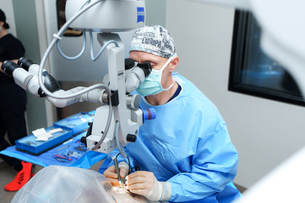
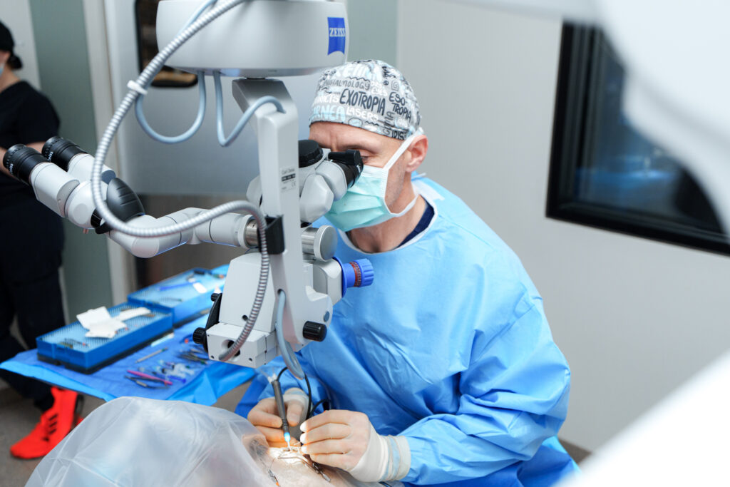
Eyelid surgery is planned in millimeters. Both undercorrection and overcorrection can create problems, and healing changes the tissues over time. Follow-up checks are important to assess the cornea, sutures, lid position, and tear film.
When to request specialist care
An eye specialist may help with complex face shape, repeat ulcers, hidden lashes, or failed past surgery. Large lumps and repair near the eye are other reasons. Read when specialist eye care may help. If haze or severe pain starts fast, review the pet eye emergency signs and get prompt care.
For COVE’s ophthalmology team, exam and surgical capabilities, Central Ohio locations, and verified contact information, visit the COVE Ophthalmology service page. An online description cannot determine whether an eyelid change is painful, whether the cornea is ulcerated, or what procedure is appropriate.