A corneal ulcer is a wound on the clear front of the eye. It may start with a scratch, infection, odd lash, or dry eye. Even a small ulcer can hurt. Some ulcers become deep very fast and can put the eye at risk.
Call a vet soon if your pet squints, holds an eye closed, rubs the face, or has a cloudy spot. A deep wound, fast cloudiness, bulging tissue, or sudden loss of sight is an emergency. Do not use human drops, old medicine, or home cures. Medicine for swelling may be unsafe when an ulcer is present.
The short answer
A corneal ulcer is often painful, even when it looks small. A vet uses a bright stain to find the wound. Shallow ulcers may heal with medicine and eye protection. Deep or infected wounds may need strong care or surgery. A recheck is key because the surface can look better before it is safe.
Keep your pet from rubbing the eye. Use a cone if the vet has shown you how. Give each drop on time, and keep the bottle tip clean.
What the cornea does
The cornea is the transparent outer window that admits and bends light. It has several thin, precisely organized layers but no blood vessels in its healthy central region. That clarity is essential for vision. The cornea contains many nerve endings, so damage can be intensely uncomfortable even when the wound is hard to see.
A shallow ulcer affects the outer layer. A stromal ulcer reaches deeper support tissue. A descemetocele is so deep that only the last thin layer is left. A full break passes through the whole cornea. Deeper wounds are more urgent and are more likely to need surgery.
Symptoms of a corneal ulcer
Possible signs include:
- Squinting or holding the eye closed
- Rapid blinking, tearing, or sensitivity to light
- Redness of the white of the eye or surrounding tissues
- Pawing at the eye or rubbing the face
- Clear, mucus-like, or yellow-green discharge
- A blue, gray, white, or uneven area on the cornea
- Swollen eyelids
- A small or irregular pupil caused by inflammation inside the eye
Cats may hide, resist touch, or become less active. A pet can have a painful ulcer without dramatic discharge. If signs begin suddenly after outdoor activity, grooming, play with another animal, or trauma, contact a vet rather than waiting for the eye to improve on its own.
What causes corneal ulcers?
Common causes include scratches, plants, dirt, chemicals, infection, odd lashes, rolled lids, and dry eye. Poor blinking or weak face nerves can also play a role. Flat-faced breeds may face more risk. Their eyes are often more open, and their lids may not cover the cornea well.
Some shallow ulcers do not heal because the outer cells fail to stick to the layer below. In dogs, these wounds are often called indolent ulcers. They are also known as SCCEDs. An antibiotic drop alone does not fix the weak bond.
In cats, feline herpesvirus can contribute to recurrent corneal disease, although not every feline ulcer is caused by herpesvirus. Diagnosis and treatment need to reflect the individual cat’s exam rather than a presumed cause.
How vets diagnose an ulcer
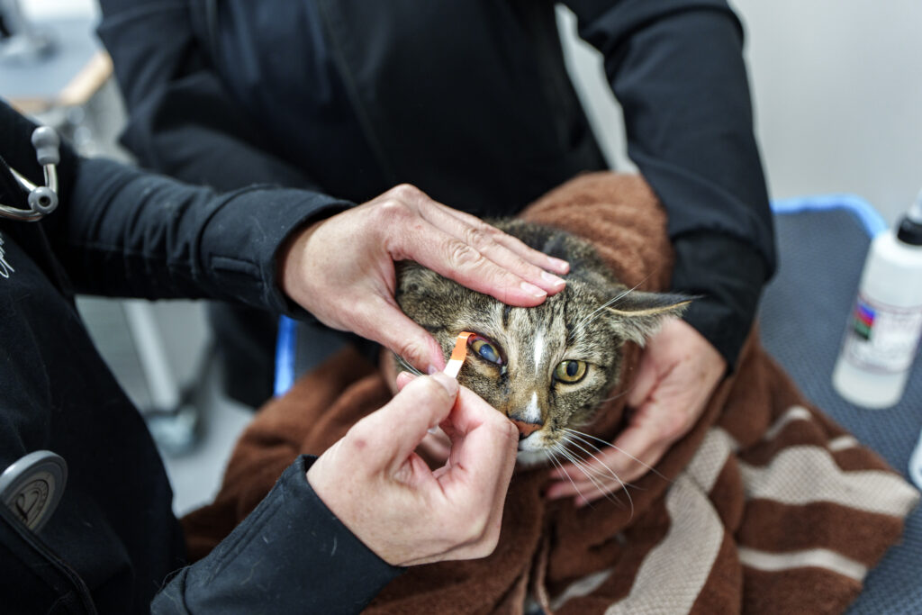
Fluorescein is a colored dye that adheres to exposed corneal stroma but not to intact epithelium. A positive stain helps identify the location and approximate shape of many ulcers. Very deep defects require careful interpretation because the thinnest central area may not retain the dye.
The vet may also test tears and eye pressure. A close view helps check the lids, lashes, and trapped material. If infection or fast tissue loss is a concern, the vet may collect cells. A sample can show which germ is present and which medicine may work.
The Merck Veterinary Manual discussion of corneal disease describes the importance of ulcer depth, infection, progressive collagen breakdown, and underlying causes in treatment planning.
Treatment for superficial ulcers
A simple shallow ulcer may need medicine for germs, pain relief, and wetting drops. An Elizabethan collar stops rubbing. The cause must also be fixed. Most pets need a recheck and another stain. This confirms that the surface has healed.
Never stop treatment solely because the eye looks better. Surface cells can cover part of the wound while the cornea remains vulnerable. Likewise, do not continue medicine longer than prescribed without follow-up. Eye conditions and medicine safety can change as healing progresses.
When a superficial ulcer does not heal
A wound that will not heal may need loose cells removed. A small tool can help new cells stick. The method may use gentle cleaning, a diamond burr, or another eye procedure. Odd lashes, rolled lids, dry eye, poor closure, and nerve problems also need care.
Repeatedly prescribing drops without reassessing ulcer depth, tear production, eyelid anatomy, and healing behavior can allow the underlying cause to persist. Referral to a veterinary ophthalmologist may be recommended for recurring, unusual, or nonhealing ulcers.
Deep, infected, and melting ulcers
Some germs and immune cells release enzymes that break down the cornea. The tissue may look soft or jelly-like. This is often called a “melting” ulcer. Tissue can be lost fast. The pet may need eye medicine around the clock, pain relief, close rechecks, and care based on culture results.
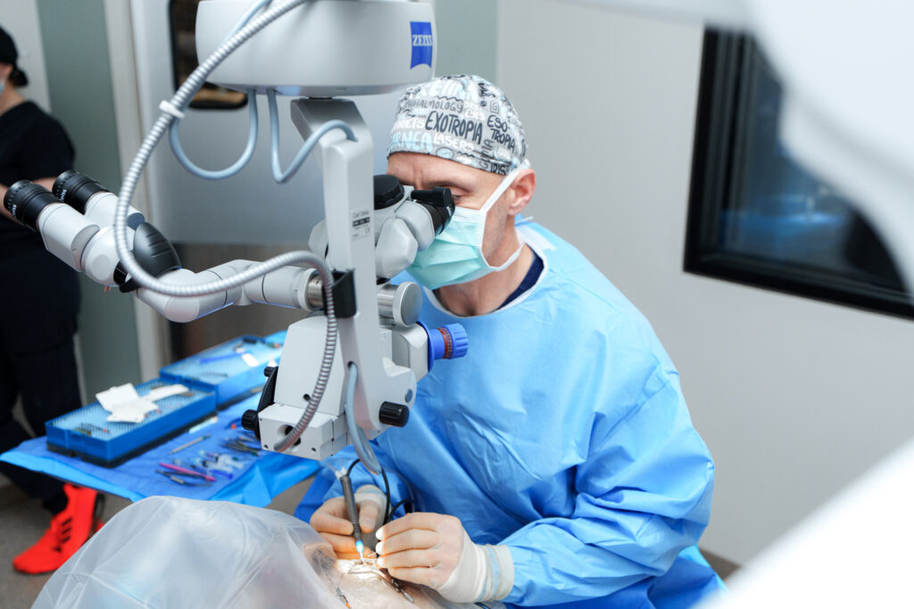
A deep ulcer, descemetocele, or perforation may require surgery to support or seal the cornea. Procedures can include a conjunctival graft, corneoscleral transposition, biologic graft material, or other reconstructive technique selected for the wound. The immediate priorities are to preserve the eye, control infection and pain, and protect whatever vision remains. A visual outcome cannot be guaranteed.
Recovery and home care
Medicine may be needed often, mainly for infected or melting ulcers. Wash your hands first. Do not touch the bottle tip to the eye. Leave the advised time between drops. Keep the collar on if told to do so. Avoid dust, branches, rough play, and rubbing.
Contact the veterinary team immediately if squinting increases, the cornea becomes more cloudy or uneven, discharge worsens, the eye changes shape, medicine cannot be given, or your pet seems ill. Rechecks are essential because the eye’s appearance at home does not reliably show depth or strength of healing.
Can corneal ulcers be prevented?
Not every wound can be prevented. Still, treat dry eye and harmful lid or lash problems. Use a collar as directed. Trim sharp plants, and do not let a pet hang its head out a car window. Pets with repeat ulcers may need a plan for their body shape or long-term disease.
If an ulcer is painful, deep, infected, recurring, or not healing as expected, ask whether specialist exam would help. Visit COVE’s Veterinary Ophthalmology service page for eye-care capabilities, team information, Central Ohio locations, and verified contact details. The guide to red, squinting, and discharging eyes in dogs explains why a surface injury can resemble other diseases. For sudden injuries, vision changes, or severe pain, review the pet eye emergency warning signs and seek immediate veterinary guidance.